

Effects of senescent secretory phenotype acquisition on human retinal pigment epithelial stem cells
- PMID: 30444724
- PMCID: PMC6286820
- DOI: 10.18632/aging.101624
Effects of senescent secretory phenotype acquisition on human retinal pigment epithelial stem cells
Abstract
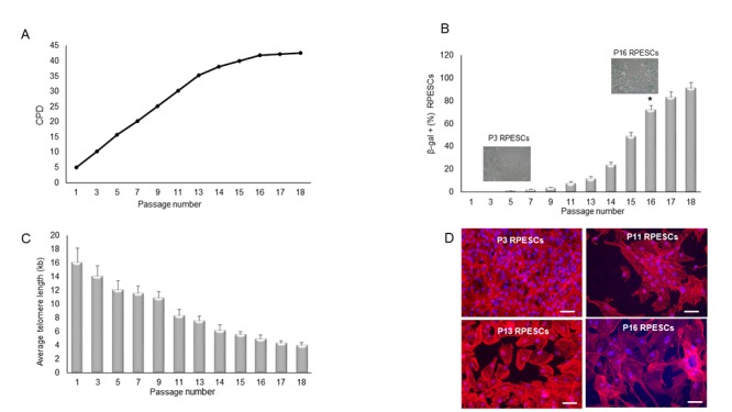
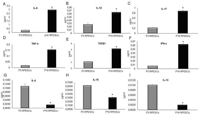
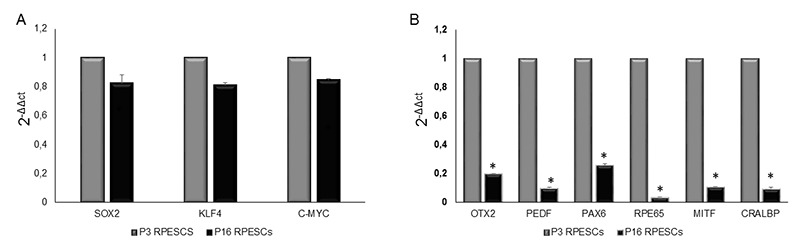
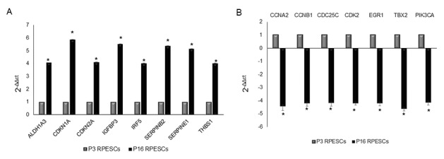
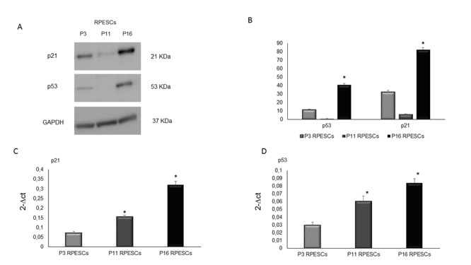
Regenerative medicine approaches based on mesenchymal stem cells (MSCs) are being investigated to treat several aging-associated diseases, including age-related macular degeneration (AMD). Loss of retinal pigment epithelium (RPE) cells occurs early in AMD, and their transplant has the potential to slow disease progression.The human RPE contains a subpopulation of cells - adult RPE stem cells (RPESCs) - that are capable of self-renewal and of differentiating into RPE cells in vitro. However, age-related MSC changes involve loss of function and acquisition of a senescence-associated secretory phenotype (SASP), which can contribute to the maintenance of a chronic state of low-grade inflammation in tissues and organs.In a previous study we isolated, characterized, and differentiated RPESCs. Here, we induced replicative senescence in RPESCs and tested their acquisition of the senescence phenotype and the SASP as well as the differentiation ability of young and senescent RPESCs.Senescent RPESCs showed a significantly reduced proliferation ability, high senescence-associated β-galactosidase activity, and SASP acquisition. RPE-specific genes were downregulated and p21 and p53 protein expression was upregulated.These findings document the effects of senescence and SASP acquisition on RPESC differentiation ability and highlight the need for a greater understanding of their role in AMD pathogenesis.
Keywords: AMD; RPESCs; age-related diseases; inflammation; senescence.
Conflict of interest statement
Figures
Similar articles
-
Oxidative stress in retinal pigment epithelium impairs stem cells: a vicious cycle in age-related macular degeneration.Mol Cell Biochem. 2022 Jan;477(1):67-77. doi: 10.1007/s11010-021-04258-3. Epub 2021 Sep 17. Mol Cell Biochem. 2022. PMID: 34535868
-
Mechanisms of RPE senescence and potential role of αB crystallin peptide as a senolytic agent in experimental AMD.Exp Eye Res. 2022 Feb;215:108918. doi: 10.1016/j.exer.2021.108918. Epub 2022 Jan 2. Exp Eye Res. 2022. PMID: 34986369 Free PMC article.
-
Reversed Senescence of Retinal Pigment Epithelial Cell by Coculture With Embryonic Stem Cell via the TGFβ and PI3K Pathways.Front Cell Dev Biol. 2020 Nov 26;8:588050. doi: 10.3389/fcell.2020.588050. eCollection 2020. Front Cell Dev Biol. 2020. PMID: 33324644 Free PMC article.
-
Senescence in the pathogenesis of age-related macular degeneration.Cell Mol Life Sci. 2020 Mar;77(5):789-805. doi: 10.1007/s00018-019-03420-x. Epub 2020 Jan 2. Cell Mol Life Sci. 2020. PMID: 31897543 Free PMC article. Review.
-
Cellular senescence in the aging retina and developments of senotherapies for age-related macular degeneration.J Neuroinflammation. 2021 Jan 22;18(1):32. doi: 10.1186/s12974-021-02088-0. J Neuroinflammation. 2021. PMID: 33482879 Free PMC article. Review.
Cited by
-
Analysis of the senescence secretome during zebrafish retina regeneration.Front Aging. 2025 Apr 16;6:1569422. doi: 10.3389/fragi.2025.1569422. eCollection 2025. Front Aging. 2025. PMID: 40308558 Free PMC article.
-
A Multi-Omics Approach Identifies Key Regulatory Pathways Induced by Long-Term Zinc Supplementation in Human Primary Retinal Pigment Epithelium.Nutrients. 2020 Oct 6;12(10):3051. doi: 10.3390/nu12103051. Nutrients. 2020. PMID: 33036197 Free PMC article.
-
Age-Related Macular Degeneration: A Disease of Cellular Senescence and Dysregulated Immune Homeostasis.Clin Interv Aging. 2024 May 23;19:939-951. doi: 10.2147/CIA.S463297. eCollection 2024. Clin Interv Aging. 2024. PMID: 38807637 Free PMC article. Review.
-
IDO1-mediated kynurenine production inhibits IGFBP5 signaling to promote 5-fluorouracil-induced senescence escape and chemoresistance in colorectal cancer.Am J Cancer Res. 2024 Sep 25;14(9):4551-4566. doi: 10.62347/XTRC3347. eCollection 2024. Am J Cancer Res. 2024. PMID: 39417170 Free PMC article.
-
Protein Acetylation in Age-Related Macular Degeneration: Mechanisms, Roles, and Therapeutic Perspectives.Invest Ophthalmol Vis Sci. 2025 May 1;66(5):30. doi: 10.1167/iovs.66.5.30. Invest Ophthalmol Vis Sci. 2025. PMID: 40402519 Free PMC article. Review.
References
-
- Hyttinen JM, Błasiak J, Niittykoski M, Kinnunen K, Kauppinen A, Salminen A, Kaarniranta K. DNA damage response and autophagy in the degeneration of retinal pigment epithelial cells-Implications for age-related macular degeneration (AMD). Ageing Res Rev. 2017; 36:64–77. 10.1016/j.arr.2017.03.006 - DOI - PubMed
MeSH terms
Substances
LinkOut - more resources
Full Text Sources
Medical
Research Materials
Miscellaneous
