. 2018 Nov 16;14(11):e1006589.
doi: 10.1371/journal.pcbi.1006589. eCollection 2018 Nov.

Towards a molecular basis of ubiquitin signaling: A dual-scale simulation study of ubiquitin dimers

Affiliations

Towards a molecular basis of ubiquitin signaling: A dual-scale simulation study of ubiquitin dimers

Andrej Berg et al. PLoS Comput Biol. .

Abstract

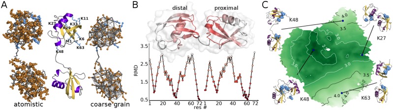
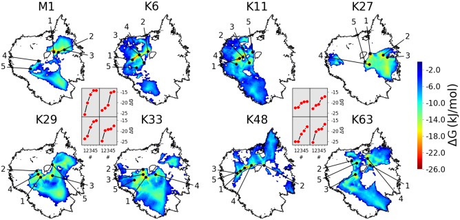
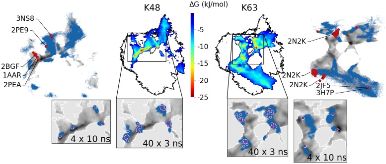
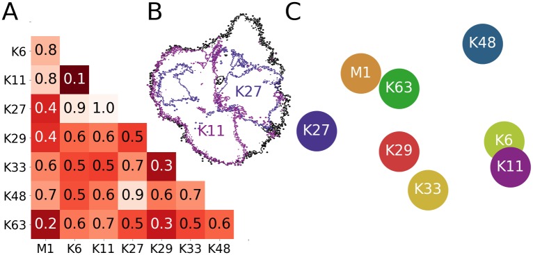
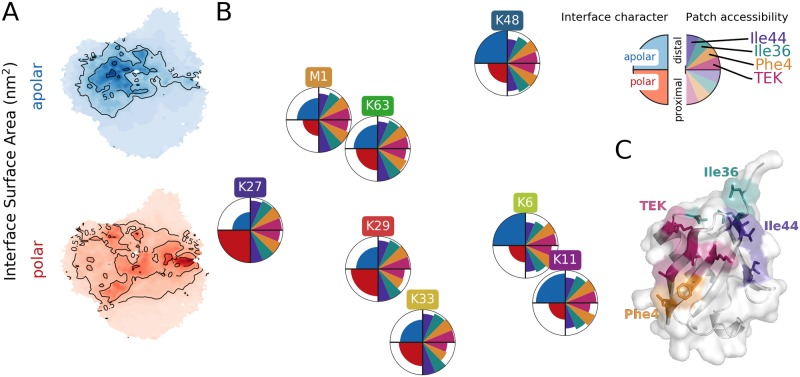
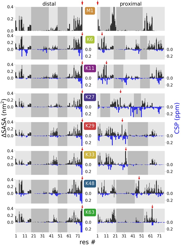
Covalent modification of proteins by ubiquitin or ubiquitin chains is one of the most prevalent post-translational modifications in eukaryotes. Different types of ubiquitin chains are assumed to selectively signal respectively modified proteins for different fates. In support of this hypothesis, structural studies have shown that the eight possible ubiquitin dimers adopt different conformations. However, at least in some cases, these structures cannot sufficiently explain the molecular basis of the selective signaling mechanisms. This indicates that the available structures represent only a few distinct conformations within the entire conformational space adopted by a ubiquitin dimer. Here, molecular simulations on different levels of resolution can complement the structural information. We have combined exhaustive coarse grained and atomistic simulations of all eight possible ubiquitin dimers with a suitable dimensionality reduction technique and a new method to characterize protein-protein interfaces and the conformational landscape of protein conjugates. We found that ubiquitin dimers exhibit characteristic linkage type-dependent properties in solution, such as interface stability and the character of contacts between the subunits, which can be directly correlated with experimentally observed linkage-specific properties.


Conflict of interest statement

The authors have declared that no competing interests exist.

Figures

Fig 1
Fig 1. Ubiquitin dimers.
(A) Proximal subunit linked via K48 to C terminus of distal subunit. Left: atomistic representation (gray: backbone atoms, brown: side-chains atoms, blue: lysine residues, i.e. alternative linkage positions, on proximal chain). Middle: cartoon representation of secondary structure. Right: coarse grained (CG) representation including supportive elastic network. (B) Residue-wise minimum distance (RMD in nm) for a K48-linked diUb structure. Residues are colored according to RMD (nm) values shown in diagram. (C) Sketch-map projection of all CG simulation structures (all linkage types). Coloring according to center of geometry distance between Ub subunits. Labels according to the linkage type illustrate conformational characteristics of certain map regions.
Fig 2
Fig 2. Conformational landscapes of diUb.
Sketch-map projected RMD data. Black circular shapes: rim of combined landscape of all linkage types (Fig 1C). Colored maps: Boltzmann-inverted probability distributions from CG simulations of individual linkage types. Black points: five lowest free-energy minima (respective energies shown in insets).
Fig 3
Fig 3. Comparison of atomistic and CG simulations.
Colored heatmaps in center: CG energy landscapes of K48- and K63-linked diUb (as in Fig 2). Outer panels: CG data in gray scale with data from atomistic simulations from open initial conformations superimposed as blue dots. Red points: experimental PDB structures. Bottom insets: Zoom with data from atomistic simulations started from back-mapped CG structures as blue dots (violet points: initial structures).
Fig 4
Fig 4. Comparison of 2D projections.
(A) Normalized earth mover distances (EMDs) between all projections in Fig 2 (i.e. all linker types). (B) Comparison of conformational landscapes to illustrate the EMD metric: all CG simulations (black); K11- and K27-linked diUb (pink and violet; EMD of 1.0). (C) 2D arrangement of linkage types based on their pairwise EMDs (i.e. according to (dis)similarity).
Fig 5
Fig 5. Characteristics of contact interface between Ub subunits from CG simulations.
(A) Mean interface surface area between distal and proximal chain on sketch-map projection divided in polar and apolar parts. (B) Circles positioned as in Fig 4C. Left half of circles show interface character, e.g. K27 has the most polar, K6 and K48 the most apolar interface. Right half of circles shows the accessibility of four known interaction patches on the distal and proximal chain. (C) Interaction patches of Ub.
Fig 6
Fig 6. Residue-wise ΔSASA values (black bars, top) compared to NMR data (blue bars, bottom).
ΔSASA calculated from CG simulations show the mean loss of accessibility for each residue and therefore the extent of interaction inside the dimer (error bars were estimated from the variance of ΔSASA for the 12 independent simulations for each linkage type). Chemical shift differences (CSP) between the distal (left) or proximal (right) Ub units in the dimer and the monomeric Ub from [15] made available by D. Fushman (no experimental data is available for M1-linked dimers). Shaded areas show regions with low ΔSASA values indicating potential interaction faces for diUb recognition. Red arrows indicate linkage position. Note that high CSP values can in part also originate from chemical modification of the lysine side chain on the proximal subunit and the C-terminus on the distal subunit [15].

Similar articles

Cited by

References

    1. Hershko A, Ciechanover A. The Ubiquitin System. Annu Rev Biochem. 1998;67(1):425–479. 10.1146/annurev.biochem.67.1.425 - DOI - PubMed
    1. Kravtsova-Ivantsiv Y, Sommer T, Ciechanover A. The Lysine48-Based Polyubiquitin Chain Proteasomal Signal: Not a Single Child Anymore. Angew Chem Int Ed. 2012;52(1):192–198. 10.1002/anie.201205656 - DOI - PubMed
    1. Swatek KN, Komander D. Ubiquitin Modifications. Cell Res. 2016;26(4):399–422. 10.1038/cr.2016.39 - DOI - PMC - PubMed
    1. Komander D, Rape M. The Ubiquitin Code. Annu Rev Biochem. 2012;81(1):203–229. 10.1146/annurev-biochem-060310-170328 - DOI - PubMed
    1. Dikic I, Wakatsuki S, Walters KJ. Ubiquitin-binding Domains—from Structures to Functions. Nat Rev Mol Cell Biol. 2009;10(10):659–671. 10.1038/nrm2767 - DOI - PMC - PubMed

Publication types