Comparative Study
. 2018 Nov 16;13(11):e0201935.
doi: 10.1371/journal.pone.0201935. eCollection 2018.

Same but different: Comparison of two system-specific molecular chaperones for the maturation of formate dehydrogenases

Affiliations
Comparative Study

Same but different: Comparison of two system-specific molecular chaperones for the maturation of formate dehydrogenases

Nadine Schwanhold et al. PLoS One. .

Abstract

The maturation of bacterial molybdoenzymes is a complex process leading to the insertion of the bulky bis-molybdopterin guanine dinucleotide (bis-MGD) cofactor into the apo-enzyme. Most molybdoenzymes were shown to contain a specific chaperone for the insertion of the bis-MGD cofactor. Formate dehydrogenases (FDH) together with their molecular chaperone partner seem to display an exception to this specificity rule, since the chaperone FdhD has been proven to be involved in the maturation of all three FDH enzymes present in Escherichia coli. Multiple roles have been suggested for FdhD-like chaperones in the past, including the involvement in a sulfur transfer reaction from the l-cysteine desulfurase IscS to bis-MGD by the action of two cysteine residues present in a conserved CXXC motif of the chaperones. However, in this study we show by phylogenetic analyses that the CXXC motif is not conserved among FdhD-like chaperones. We compared in detail the FdhD-like homologues from Rhodobacter capsulatus and E. coli and show that their roles in the maturation of FDH enzymes from different subgroups can be exchanged. We reveal that bis-MGD-binding is a common characteristic of FdhD-like proteins and that the cofactor is bound with a sulfido-ligand at the molybdenum atom to the chaperone. Generally, we reveal that the cysteine residues in the motif CXXC of the chaperone are not essential for the production of active FDH enzymes.


Conflict of interest statement

The authors have declared that no competing interests exist.

Figures

Fig 1
Fig 1. Phylogenetic tree of FdhD-like proteins.
Protein phylogeny of FdsC/FdhD homologues based on a full length multiple sequence alignment by Muscle [50]. The tree was constructed using the Maximum Likelihood method based on the Dayhoff matrix based model [51][57]. The tree is drawn to scale, with branch lengths in the same units as those of the evolutionary distances used to infer the phylogenetic tree. The evolutionary distances were computed using the Dayhoff matrix based method and are in the units of the number of amino acid substitutions per site [52]. The scale bar indicates 0.2 substitutions per site. Numbers near branches indicate the bootstrap proportion for 100 replicas using the same method. The analysis involved 41 amino acid sequences. All positions containing gaps and missing data were eliminated. There was a total of 174 positions in the final dataset. Evolutionary analyses were conducted in MEGA7 [53]. Domains and classes of prokaryotes are marked in blue: α- Proteobacteria, β-Proteobacteria, γ-Proteobacteria, δ-Proteobacteria, ε-Proteobacteria. There are three main branches: group I, II and III. FdsC/FdhD homologues in group I and II harbor the conserved CXXC-motif, which is not present in group III (♦ - CC-motif, □ - only second cysteine, ■ - CXXXCXC-motif, ◙ - no cysteine). Existing FDHs are listed as described in the box on the left and based on genomic analysis using www.ncbi.nlm.nih.gov or * img.jgi.doe.gov. If already described in literature, the corresponding active site metal (Mo or W) is displayed.
Fig 2
Fig 2. Amino acid sequence alignment of E. coli FdhD and R. capsulatus FdsC.
The amino acid sequence alignment was generated using ClustalW. Identical amino acids and similar amino acids are marked with a black and a grey box, respectively. The conserved cysteine residues are labelled with red arrows. The blue bar over the alignment indicates the N-terminal domain (blue), the linker region (yellow) and the C-terminal domain (green) based on the structure of FdhD (PDB: 4pde) [19].
Fig 3
Fig 3. Characterization of cysteine variants of FdsC and FdhD.
FdsC and FdhD and their cysteine variants were expressed in E. coli ΔfdhD strain and purified by Ni-NTA chromatography as described for FdsC previously [21]. A) 15 μg of purified FdsC, FdsC-C104A, FdsC-C107A and FdsC-C104/C107A were separated by SDS-PAGE. B) 15 μg of purified FdhD, FdhD-C121A, FdhD-C124A and FdhD-C121/C124A were separated by SDS-PAGE. C) 200–300 μM of protein was treated with I2/KI-HCl to isolate Form A-GMP as described previously [47]. Form A-GMP was separated by HPLC. The corresponding peak area was normalized for protein concentration. The results represent the mean values from three independent measurements (±S.D.).
Fig 4
Fig 4. Reconstitution of E. coli apo-TMAO reductase using FdsC or FdhD as bis-MGD source.
50 μM of each purified chaperone variant was incubated with apo-TMAO reductase (8 μM) to reconstitute enzyme activity. After 2 hours TMAO reductase activity was measured under anaerobic conditions following the oxidation of reduced benzyl viologen at 600 nm in the presence of 0.1 μM TMAO. Kinetic Data are mean values from three independent measurements (±S.D.).
Fig 5
Fig 5. Characterization of E. coli apo-TMAO reductase reconstituted with FdsC or FdhD.
For the reconstitution of enzyme activity, 50 μM of each FdhD, FdsC or their variants was incubated with apo-TMAO reductase (8 μM) in a total volume of 8 ml. After 7 hours, the samples were concentrated to 500 μl before size exclusion chromatography. The results represent the mean values from three independent measurements (±S.D.). A) Relative Form A-GMP content (MGD in LU*s per mg TMAO reductase) of 3 μM TMAO reductase was analyzed by HPLC. B) Activity of the fraction containing TMAO reductase was measured under anaerobic conditions following the oxidation of reduced benzyl viologen at 600 nm in the presence of 0.1 μM TMAO. TMAO reductase activity was normalized for the MGD content.
Fig 6
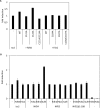
Fig 6. Influence of FdsC and FdhD on FDH activity.
A) R. capsulatus (FdsGBA)2 was expressed in ΔfdhD strain in the presence of FdhD WT (pNB14), FdhD-C121A (pNB15), FdhD-C124A (pNB16), FdhD-C121A/C124A (pNB17) or FdsC WT (pTHfds03) and purified by Ni-NTA affinity chromatography as described previously [21]. R. capsulatus (FdsGBA)2 activity was detected photometrically by the increase in NADH recorded at 340 nm and 1 U/mg is defined as the reduction of 1 μmol NAD+/min/mg of enzyme at room temperature. Kinetic Data are mean values from three independent measurements (±S.D.).The enzyme expressed in the absence of FdsC or FdhD was not active (data not shown), as reported previously [21]. B) 55 ml cultures of each strain were grown anaerobically at 37°C for 16 hours in LB media in the presence of 20 μM IPTG, 10 μM sodium molybdate and antibiotic as needed. Equivalent amounts (40–60 μg) of Triton-X100 treated crude extracts were applied to each lane and separated by non-denaturing PAGE. The gels were stained with 1 mM NBT, 0.5 mM PMS and 50 mM formate in 50 mM potassium phosphate, pH 6.8. Lanes from left to right: BW25113, ΔfdoG, ΔfdhD, +pFdhD: ΔfdhD deficient strain complemented with plasmid pNB14, +pFdhD-C121A: ΔfdhD deficient strain complemented with plasmid pNB15, +pFdhD-C124A: ΔfdhD deficient strain complemented with plasmid pNB16, +pFdhD-C121A/C124A: ΔfdhD deficient strain complemented with plasmid pNB15, +pFdsC: ΔfdhD deficient strain complemented with plasmid pTHfds14, +pFdsC-C104A: ΔfdhD deficient strain complemented with plasmid pNBfds04, +pFdsC-C107A: ΔfdhD deficient strain complemented with plasmid pNBfds05, +pFdsC-C104A/C107A: ΔfdhD deficient strain complemented with plasmid pNBfds06.
Fig 7
Fig 7. Influence of FdsC and FdhD on l-cysteine desulfurase activity.
l-cysteine desulfurase activity was measured by determination of total sulfide produced [48]. A) 1 μM IscS from E. coli was mixed with either 2 μM FdhD/FdhD variants or FdsC/FdsC variants and incubated for 10 min in the presence of 1 mM l-cysteine at 30°C. The activity of IscS alone was set to 1. B) l-cysteine desulfurase IscS (1 μM) from E. coli or NifS4, NifS3 and NifS2-Δ1–188 from R. capsulatus (2 μM) were mixed in a 1:2 ratio with FdsC, FdhD or SufE, respectively and incubated for 10 min in the presence of 1 mM l-cysteine at 30°C. The fold induction of IscS, NifS4, NifS3 or NifS2-Δ1–188 activity by incubation with FdsC, FdhD or SufE is relative to the activity of the l-cysteine desulfurase alone, respectively. Kinetic Data are mean values from three independent measurements (±S.D.).
Fig 8
Fig 8. Interaction of FdhD with IscS.
Complex formation of (A) 40 μM FdhD + 20 μM IscS, (B) 40 μM FdhD-C121A/C124A + 20 μM IscS, (C) 40 μM FdsC + 20 μM IscS and (D) 40 μM FdsC-C104A/C107A + 20 μM IscS were analyzed by size exclusion chromatography on a Superdex Increase 200 column (GE Healthcare) equilibrated in 100 mM potassium phosphate buffer, pH 8.0, 100 mM NaCl, 10 mM β-mercaptoethanol. The elution of proteins was followed at 280 nm. Indicated fractions in a range of 10.5–15.5 ml (0.5 ml fractions) were analyzed for their protein content by 15%SDS−PAGE.
Fig 9
Fig 9. Model for bis-MGD sulfuration and insertion into the target enzymes by involvement of FdsC and FdhD.
FdsC and FdhC bind the bis-MGD cofactor synthesized by the Moco biosynthesis machinery and provide the sulfurated bis-MGD for the FDHs of R. capsulatus and E. coli, respectively. Both chaperones are involved in the sulfuration of bis-MGD. FdhD interacts with the L-cysteine desulfurase IscS. The corresponding L-cysteine desulfurase for R. capsulatus has not been identified to date. FdsC and FdhD are functional homologues since the can replace each other in the maturation of the respective FDH.

Similar articles

Cited by

References

    1. Leimkühler S, Iobbi-Nivol C. Bacterial molybdoenzymes: old enzymes for new purposes. FEMS microbiology reviews. 2016;40(1):1–18. Epub 2015/10/16. 10.1093/femsre/fuv043 . - DOI - PubMed
    1. Hille R. The mononuclear molybdenum enzymes. Chemical Rev. 1996;96:2757–816. - PubMed
    1. Palmer T, Vasishta A, Whitty PW, Boxer DH. Isolation of protein FA, a product of the mob locus required for molybdenum cofactor biosynthesis in Escherichia coli. Eur J Biochem. 1994;222:687–92. - PubMed
    1. Yokoyama K, Leimkühler S. The role of FeS clusters for molybdenum cofactor biosynthesis and molybdoenzymes in bacteria. Biochim Biophys Acta. 2014. Epub 2014/10/01. 10.1016/j.bbamcr.2014.09.021 . - DOI - PMC - PubMed
    1. Kisker C, Schindelin H, Rees DC. Molybdenum-cofactor-containing enzymes: structure and mechanism. Ann Rev Biochem. 1997;66:233–67. 10.1146/annurev.biochem.66.1.233 - DOI - PubMed

Publication types

MeSH terms