

Efficacy of leukemia inhibitory factor as a therapeutic for permanent large vessel stroke differs among aged male and female rats
- PMID: 30445025
- PMCID: PMC6814304
- DOI: 10.1016/j.brainres.2018.11.017
Efficacy of leukemia inhibitory factor as a therapeutic for permanent large vessel stroke differs among aged male and female rats
Abstract
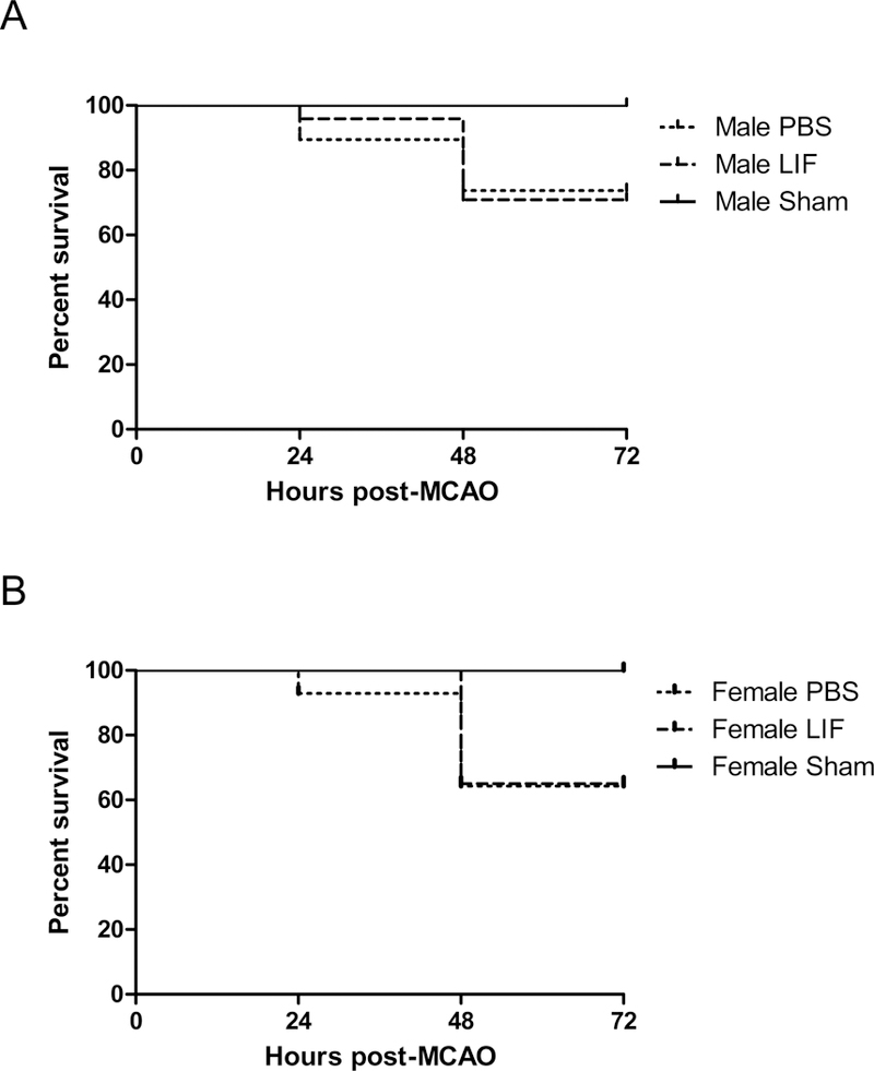
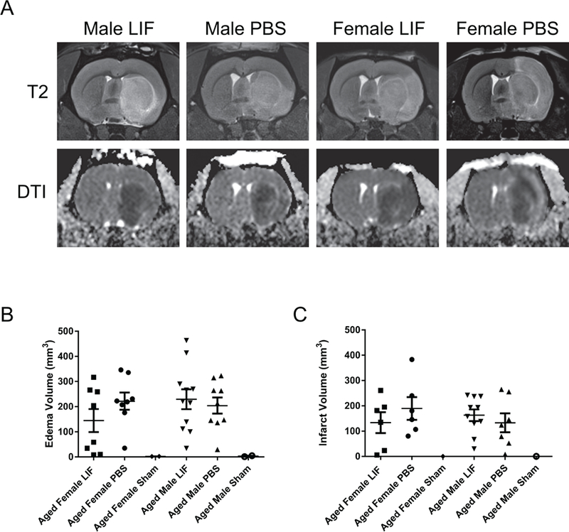
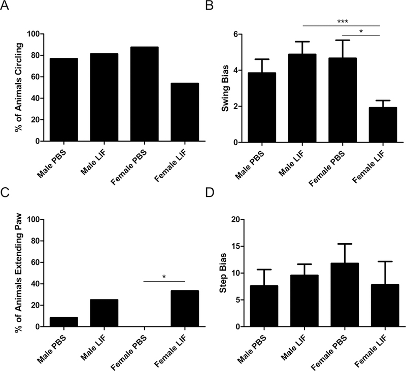
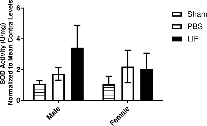
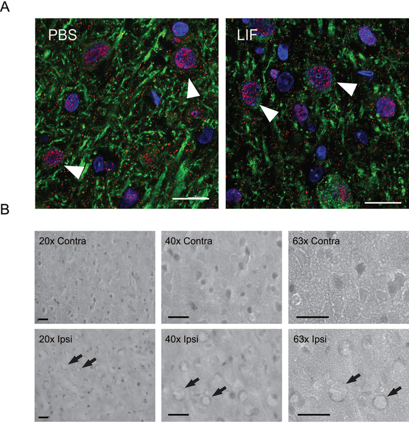
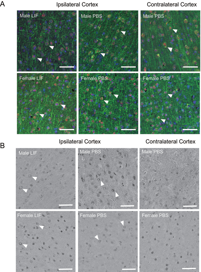
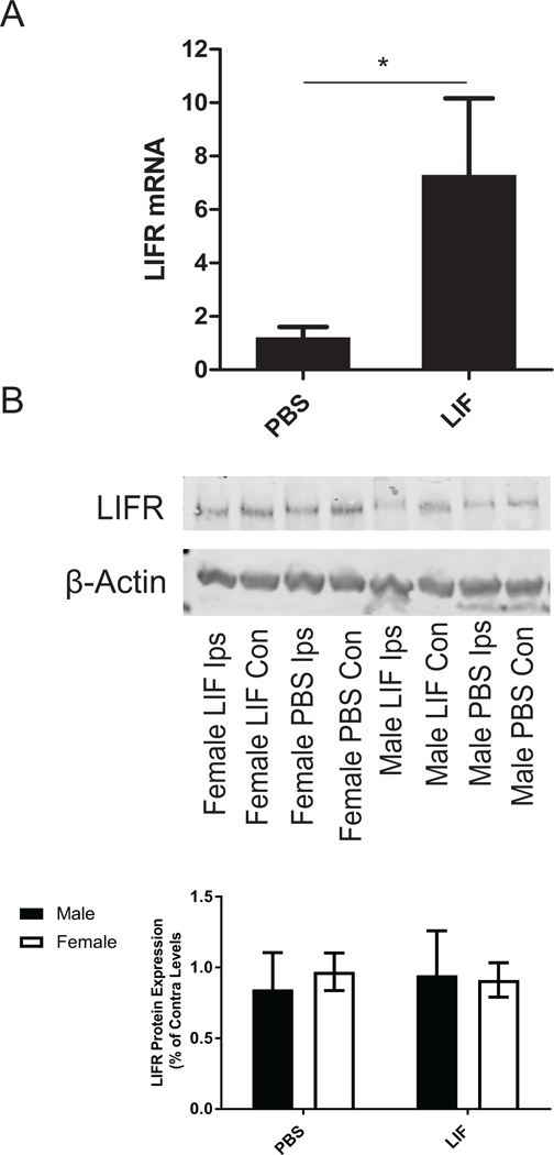
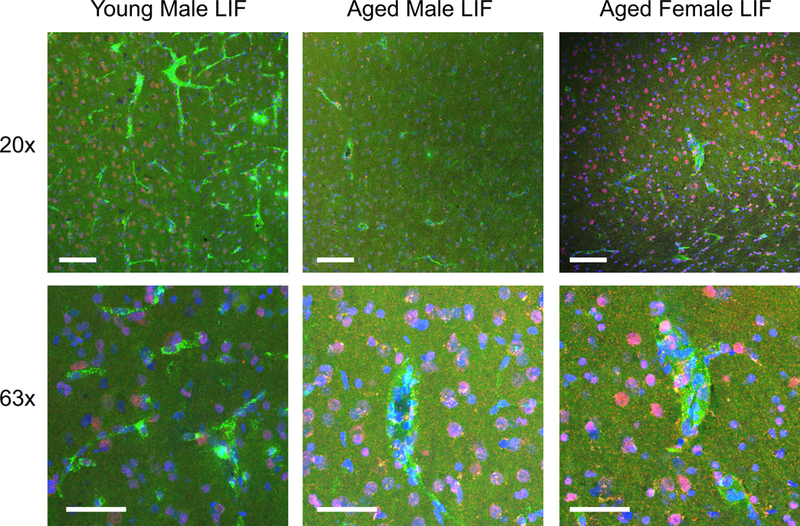
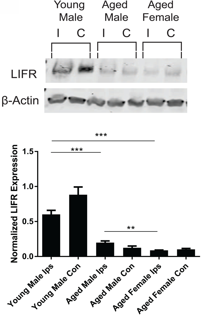
Preclinical studies using rodent models of stroke have had difficulty in translating their results to human patients. One possible factor behind this inability is the lack of studies utilizing aged rodents of both sexes. Previously, this lab showed that leukemia inhibitory factor (LIF) promoted recovery after stroke through antioxidant enzyme upregulation. This study examined whether LIF promotes neuroprotection in aged rats of both sexes. LIF did not reduce tissue damage in aged animals, but LIF-treated female rats showed partial motor skill recovery. The LIF receptor (LIFR) showed membrane localization in young male and aged rats of both sexes after stroke. Although LIF increased neuronal LIFR expression in vitro, it did not increase LIFR in the aged brain. Levels of LIFR protein in brain tissue were significantly downregulated between young males and aged males/females at 72 h after stroke. These results demonstrated that low LIFR expression reduces the neuroprotective efficacy of LIF in aged rodents of both sexes. Furthermore, the ability of LIF to promote motor improvement is dependent upon sex in aged rodents.
Keywords: Aging; Cytokine; Ischemia; Neuroprotection; Stroke.
Copyright © 2018 The Authors. Published by Elsevier B.V. All rights reserved.
Conflict of interest statement
Declarations of Interest
The authors have no interests to disclose.
Figures
Similar articles
-
Activation of cytokine signaling through leukemia inhibitory factor receptor (LIFR)/gp130 attenuates ischemic brain injury in rats.J Cereb Blood Flow Metab. 2005 Jun;25(6):685-93. doi: 10.1038/sj.jcbfm.9600061. J Cereb Blood Flow Metab. 2005. PMID: 15716858
-
Neuroprotective activity of leukemia inhibitory factor is relayed through myeloid zinc finger-1 in a rat model of stroke.Metab Brain Dis. 2019 Apr;34(2):631-640. doi: 10.1007/s11011-018-0376-2. Epub 2019 Jan 5. Metab Brain Dis. 2019. PMID: 30612292 Free PMC article.
-
Differential stimulation-induced receptor localization in lipid rafts for interleukin-6 family cytokines signaling through the gp130/leukemia inhibitory factor receptor complex.J Neurochem. 2007 May;101(3):782-93. doi: 10.1111/j.1471-4159.2007.04471.x. J Neurochem. 2007. PMID: 17448148
-
Roles of leukemia inhibitory factor receptor in cancer.Int J Cancer. 2025 Jan 15;156(2):262-273. doi: 10.1002/ijc.35157. Epub 2024 Sep 15. Int J Cancer. 2025. PMID: 39279155 Review.
-
Leukemia inhibitory factor and human embryo implantation.Ann N Y Acad Sci. 2004 Dec;1034:176-83. doi: 10.1196/annals.1335.020. Ann N Y Acad Sci. 2004. PMID: 15731310 Review.
Cited by
-
Insulin-Like Growth Factor-1 Is Neuroprotective in Aged Rats With Ischemic Stroke.Front Aging Neurosci. 2019 Dec 11;11:349. doi: 10.3389/fnagi.2019.00349. eCollection 2019. Front Aging Neurosci. 2019. PMID: 31920629 Free PMC article.
-
Sex- and Tissue-Specific Effects of Leukemia Inhibitory Factor on Mitochondrial Bioenergetics Following Ischemic Stroke.Biomolecules. 2025 May 20;15(5):738. doi: 10.3390/biom15050738. Biomolecules. 2025. PMID: 40427631 Free PMC article.
-
Translating concepts of neural repair after stroke: Structural and functional targets for recovery.Restor Neurol Neurosci. 2020;38(1):67-92. doi: 10.3233/RNN-190978. Restor Neurol Neurosci. 2020. PMID: 31929129 Free PMC article. Review.
-
The Poststroke Peripheral Immune Response Is Differentially Regulated by Leukemia Inhibitory Factor in Aged Male and Female Rodents.Oxid Med Cell Longev. 2020 Dec 10;2020:8880244. doi: 10.1155/2020/8880244. eCollection 2020. Oxid Med Cell Longev. 2020. PMID: 33376583 Free PMC article.
-
Leukemia Inhibitory Factor Receptor Is Involved in Apoptosis in Rat Astrocytes Exposed to Oxygen-Glucose Deprivation.Biomed Res Int. 2019 Feb 27;2019:1613820. doi: 10.1155/2019/1613820. eCollection 2019. Biomed Res Int. 2019. PMID: 30937308 Free PMC article.
References
-
- Ajmo CT Jr., Vernon DO, Collier L, Pennypacker KR, Cuevas J, 2006. Sigma receptor activation reduces infarct size at 24 hours after permanent middle cerebral artery occlusion in rats. Curr Neurovasc Res 3, 89–98. - PubMed
-
- Alkayed NJ, Harukuni I, Kimes AS, London ED, Traystman RJ, Hurn PD, 1998. Gender-linked brain injury in experimental stroke. Stroke 29, 159–166. - PubMed
-
- Appelros P, Stegmayr B, Terént A, 2009. Sex differences in stroke epidemiology: a systematic review. Stroke 40, 1082–1090. - PubMed
-
- Azari MF, Galle A, Lopes EC, Kurek J, Cheema SS, 2001. Leukemia inhibitory factor by systemic administration rescues spinal motor neurons in the SOD1 G93A murine model of familial amyotrophic lateral sclerosis. Brain Res 922, 144–147. - PubMed
-
- Azari MF, Lopes EC, Stubna C, Turner BJ, Zang D, Nicola NA, Kurek JB, Cheema SS, 2003. Behavioural and anatomical effects of systemically administered leukemia inhibitory factor in the SOD1 G93A G1H mouse model of familial amyotrophic lateral sclerosis. Brain Res 982, 92–97. - PubMed
Publication types
MeSH terms
Substances
Grants and funding
LinkOut - more resources
Full Text Sources
Medical
Miscellaneous
