

A tight balance of Karyopherin β1 expression is required in cervical cancer cells
- PMID: 30445944
- PMCID: PMC6240311
- DOI: 10.1186/s12885-018-5044-8
A tight balance of Karyopherin β1 expression is required in cervical cancer cells
Abstract
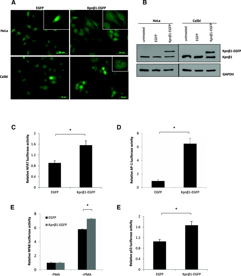
Background: Karyopherin β1 (Kpnβ1) is the main nuclear import protein involved in the transport of cargoes from the cytoplasm into the cell nucleus. Previous research has found Kpnβ1 to be significantly overexpressed in cervical cancer and other cancer tissues, and further studies showed that inhibition of Kpnβ1 expression by siRNA resulted in cancer cell death, while non-cancer cells were minimally affected. These results suggest that Kpnβ1 has potential as an anticancer therapeutic target, thus warranting further research into the association between Kpnβ1 expression and cancer progression. Here, the biological effects associated with Kpnβ1 overexpression were investigated in order to further elucidate the relationship between Kpnβ1 and the cancer phenotype.
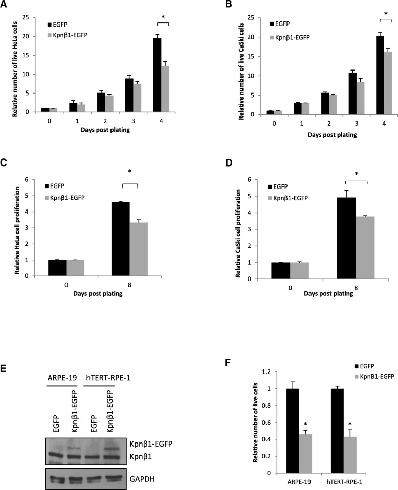
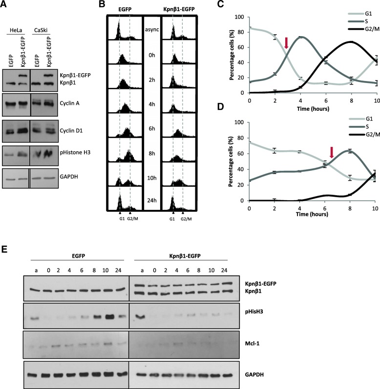
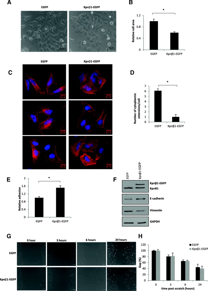
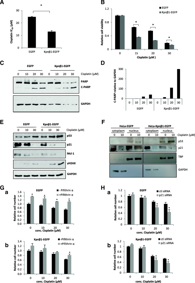
Methods: To evaluate the effect of Kpnβ1 overexpression on cell biology, cell proliferation, cell cycle, cell morphology and cell adhesion assays were performed. To determine whether Kpnβ1 overexpression influences cell sensitivity to chemotherapeutic agents like Cisplatin, cell viability assays were performed. Expression levels of key proteins were analysed by Western blot analysis.
Results: Our data revealed that Kpnβ1 overexpression, above that which was already detected in cancer cells, resulted in reduced proliferation of cervical cancer cells. Likewise, normal epithelial cells showed reduced proliferation after Kpnβ1 overxpression. Reduced cancer cell proliferation was associated with a delay in cell cycle progression, as well as changes in the morphology and adhesion properties of cells. Additionally, Kpnβ1 overexpressing HeLa cells exhibited increased sensitivity to cisplatin, as shown by decreased cell viability and increased apoptosis, where p53 and p21 inhibition reduced and enhanced cell sensitivity to Cisplatin, respectively.
Conclusions: Overall, our results suggest that a tight balance of Kpnβ1 expression is required for cellular function, and that perturbation of this balance results in negative effects associated with a variety of biological processes.
Keywords: Cancer biology; Karypherin β1; Overexpression.
Conflict of interest statement
Ethics approval and consent to participate
Not applicable.
Consent for publication
Not applicable.
Competing interests
The authors declare that they have no competing interests.
Publisher’s Note
Springer Nature remains neutral with regard to jurisdictional claims in published maps and institutional affiliations.
Figures
Similar articles
-
Inhibition of Kpnβ1 mediated nuclear import enhances cisplatin chemosensitivity in cervical cancer.BMC Cancer. 2021 Feb 2;21(1):106. doi: 10.1186/s12885-021-07819-3. BMC Cancer. 2021. PMID: 33530952 Free PMC article.
-
The Karyopherin proteins, Crm1 and Karyopherin beta1, are overexpressed in cervical cancer and are critical for cancer cell survival and proliferation.Int J Cancer. 2009 Apr 15;124(8):1829-40. doi: 10.1002/ijc.24146. Int J Cancer. 2009. PMID: 19117056 Free PMC article.
-
Novel small molecule inhibitor of Kpnβ1 induces cell cycle arrest and apoptosis in cancer cells.Exp Cell Res. 2021 Jul 15;404(2):112637. doi: 10.1016/j.yexcr.2021.112637. Epub 2021 May 18. Exp Cell Res. 2021. PMID: 34019908
-
The nuclear import receptor Kpnβ1 and its potential as an anticancer therapeutic target.Crit Rev Eukaryot Gene Expr. 2013;23(1):1-10. doi: 10.1615/critreveukargeneexpr.2013004845. Crit Rev Eukaryot Gene Expr. 2013. PMID: 23557333 Review.
-
KPNB1-mediated nuclear import in cancer.Eur J Pharmacol. 2023 Sep 15;955:175925. doi: 10.1016/j.ejphar.2023.175925. Epub 2023 Jul 18. Eur J Pharmacol. 2023. PMID: 37473981 Review.
Cited by
-
Inhibition of Kpnβ1 mediated nuclear import enhances cisplatin chemosensitivity in cervical cancer.BMC Cancer. 2021 Feb 2;21(1):106. doi: 10.1186/s12885-021-07819-3. BMC Cancer. 2021. PMID: 33530952 Free PMC article.
-
Recognition motifs for importin 4 [(L)PPRS(G/P)P] and importin 5 [KP(K/Y)LV] binding, identified by bio-informatic simulation and experimental in vitro validation.Comput Struct Biotechnol J. 2022 Oct 26;20:5952-5961. doi: 10.1016/j.csbj.2022.10.015. eCollection 2022. Comput Struct Biotechnol J. 2022. PMID: 36382187 Free PMC article.
-
Expression of Karyopherin Alpha 2 and Karyopherin Beta 1 Correlate with Poor Prognosis in Gastric Cancer.Oncology. 2022;100(12):685-695. doi: 10.1159/000526807. Epub 2022 Oct 21. Oncology. 2022. PMID: 36273446 Free PMC article.
-
Inhibiting Importin 4-mediated nuclear import of CEBPD enhances chemosensitivity by repression of PRKDC-driven DNA damage repair in cervical cancer.Oncogene. 2020 Aug;39(34):5633-5648. doi: 10.1038/s41388-020-1384-3. Epub 2020 Jul 13. Oncogene. 2020. PMID: 32661323 Free PMC article.
-
A mass spectrometry-based approach for the identification of Kpnβ1 binding partners in cancer cells.Sci Rep. 2022 Nov 23;12(1):20171. doi: 10.1038/s41598-022-24194-6. Sci Rep. 2022. PMID: 36418423 Free PMC article.
References
-
- Harel A, Forbes DJ. Importin beta: conducting a much larger cellular symphony. Mol Cell. 2004;16:319–330. - PubMed
MeSH terms
Substances
LinkOut - more resources
Full Text Sources
Other Literature Sources
Medical
Research Materials
Miscellaneous
