Review
. 2019 Feb;44(2):125-140.
doi: 10.1016/j.tibs.2018.10.005. Epub 2018 Nov 13.

PXE, a Mysterious Inborn Error Clarified

Affiliations
Review

PXE, a Mysterious Inborn Error Clarified

Piet Borst et al. Trends Biochem Sci. 2019 Feb.

Abstract

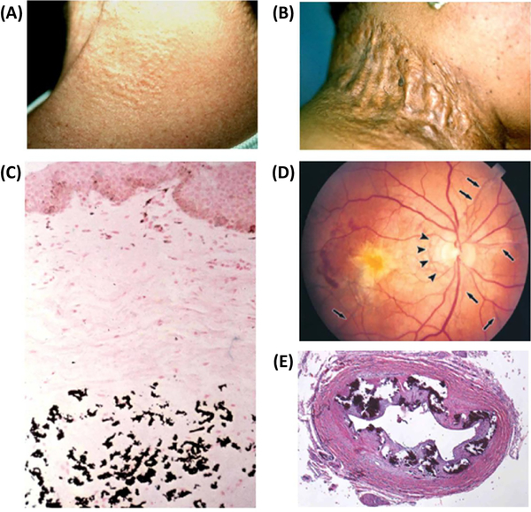
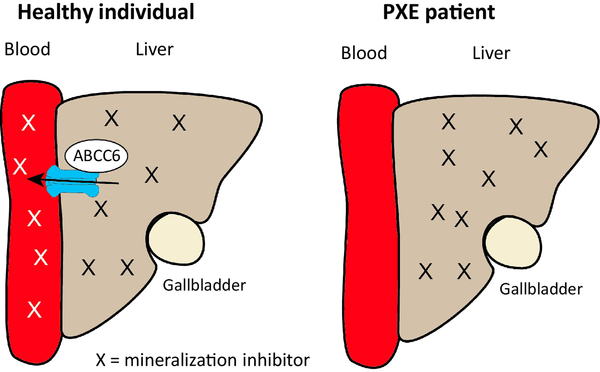
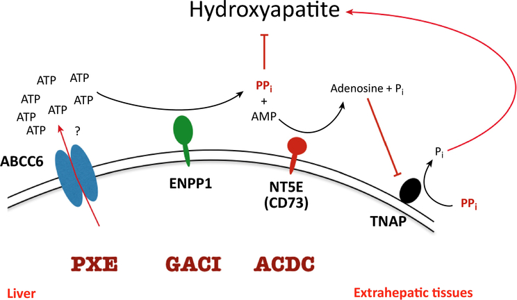
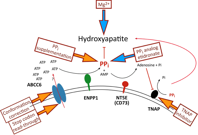
Ever since Garrod deduced the existence of inborn errors in 1901, a vast array of metabolic diseases has been identified and characterized in molecular terms. In 2018 it is difficult to imagine that there is any uncharted backyard left in the metabolic disease landscape. Nevertheless, it took until 2013 to identify the cause of a relatively frequent inborn error, pseudoxanthoma elasticum (PXE), a disorder resulting in aberrant calcification. The mechanism found was not only biochemically interesting but also points to possible new treatments for PXE, a disease that has remained untreatable. In this review we sketch the tortuous road that led to the biochemical understanding of PXE and to new ideas for treatment. We also discuss some of the controversies still haunting the field.

Keywords: ABC-transporters; ABCC6; Calcification; Pseudoxanthoma; Pyrophosphate.


Figures

Figure 1.
Figure 1.
Clinical features and histopathology of ectopic mineralization in pseudoxanthoma elasticum (PXE). (A) Characteristic early cutaneous signs of PXE consist of discrete yellowish papules at predilection sites, such as sides of the neck. (B) These early lesions coalesce into plaques of inelastic, leathery skin. (C) Staining of a parallel section with von Kossa stain reveals mineralization of the elastotic structures. (D) Characteristic ocular findings consist of angioid streaks, breaks in Bruch’s membrane behind the retina (arrows), which allow neovascularization (arrowheads) of the retina leading to loss of visual acuity and blindness. (E) Histopathology of the left renal artery in a GACI patient with ABCC6 mutations reveals extensive mineralization (Hematoxylin-Eosin stain). Adapted from Li et al (2018 Am. J. Pathol accepted for publication)
Figure 2.
Figure 2.
The “metabolic” hypothesis for PXE. ABCC6 is located in the baso-lateral membrane of the hepatocyte where it secretes a compound X into the blood circulation. X is desributed throughout the body via the circulation and hypothesised to counteract the ectopic peripheral calcification that occurs in PXE patients with non-functioning ABCC6.
Figure 3.
Figure 3.
How membrane vesicles can be used to study transport of substrates by ABC-transporters. When cells are disrupted, the plasma membrane forms two types of vesicles, inside-in vesicles in which the transporter has its normal orientation pumping substrates out; and inside-out vesicles in which the orientation is inverted, the transporter pumping substrates into the vesicle, if ATP is provided to energise the pump. Following the incubation of the vesicles with radioactive substrate and ATP, the vesicles are washed on filters and counted. The inside-in vesicles do not contribute to the reaction, as under these conditions no ATP is present inside the vesicles to support ATP hydrolysis at the ABCC6 nucleotide-binding domains.
Figure 4.
Figure 4.
Liver perfusion experiments showing that mouse liver produces large amounts of AMP and PPi, but not in mice without functional ABCC6. The very low levels of ATP in the perfusates are contributed to the rapid conversion of any ATP released into the perfusion buffer into AMP and PPi. In PXE patients reduced ABCC6-mediated ATP release results in low plasma PPi concentrations.
Figure 5.
Figure 5.
The known major mechanisms regulating plasma pyrophosphate level. Quantitatively the most important source PPi is the ABCC6-mediated ATP-release from hepatocytes. This ATP is rapidly converted into AMP and to PPi in the liver vasculature by ENPP1. The most important enzyme degrading PPi is TNAP. The activity of this enzyme is regulated by NT5E, which cleaves AMP to adenine and phosphate (Pi); the AMP generated inhibits TNAP, suppressing PPi hydrolysis. Mutations in the ABCC6, the ENPP1 and the NT5E genes cause various calcification diseases. (For more details see text).
Figure 6.
Figure 6.
Current therapeutic attempts to counteract the ectopic calcification in PXE patients. Interventions targeting different steps in the regulation of ectopic mineralization are indicated by arrows. Yellow block arrows indicate preclinical interventions, blue arrows ongoing or recently completed clinical trials. (For more details see text).

References

    1. Le Boulanger G et al. (2009) An unusual severe vascular case of pseudoxanthoma elasticum presenting as generalized arterial calcification of infancy. Am. J. Med. Genet 152, 118–123 - PubMed
    1. Uitto J et al. (2017) Insights into Pathomechanisms and Treatment Development in Heritable Ectopic Mineralization Disorders: Summary of the PXE International Biennial Research Symposium-2016. J. Invest. Dermatol 137, 790–795 - PMC - PubMed
    1. Kranenburg G et al. (2018) Etidronate for Prevention of Ectopic Mineralization in Patients With Pseudoxanthoma Elasticum. J. Am. Coll. Cardiol 71, 1117–1126 - PubMed
    1. Le Saux O et al. (2000) Mutations in a gene encoding an ABC transporter cause pseudoxanthoma elasticum. Nat. Genet 25, 223–227 - PubMed
    1. Bergen AA et al. (2000) Mutations in ABCC6 cause pseudoxanthoma elasticum. Nat. Genet 25, 228–231 - PubMed

Publication types

MeSH terms

LinkOut - more resources