. 2018 Dec 18;49(6):1062-1076.e6.
doi: 10.1016/j.immuni.2018.09.018. Epub 2018 Nov 13.

Distinct Compartmentalization of the Chemokines CXCL1 and CXCL2 and the Atypical Receptor ACKR1 Determine Discrete Stages of Neutrophil Diapedesis

Affiliations

Distinct Compartmentalization of the Chemokines CXCL1 and CXCL2 and the Atypical Receptor ACKR1 Determine Discrete Stages of Neutrophil Diapedesis

Tamara Girbl et al. Immunity. .

Abstract

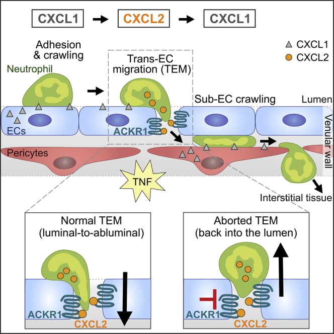
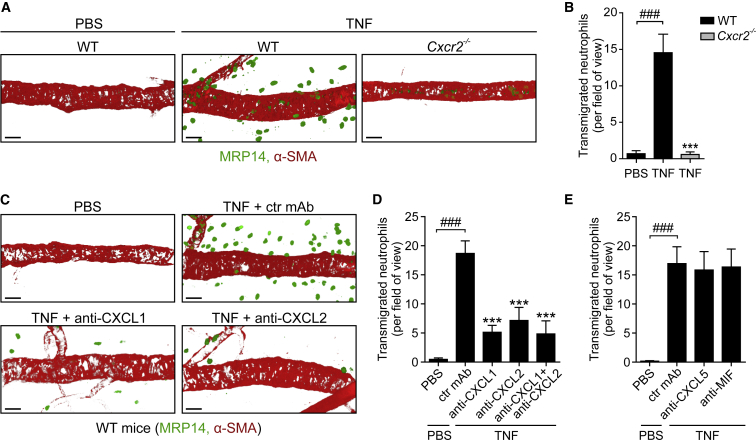
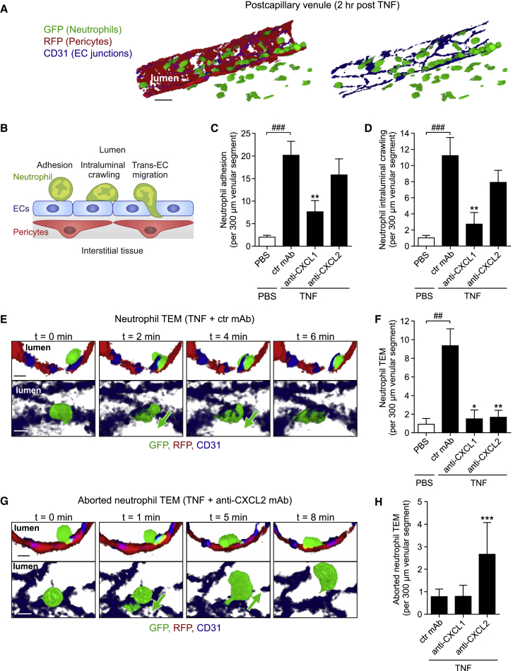
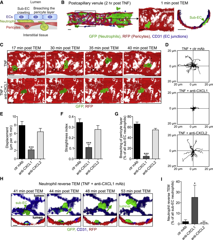
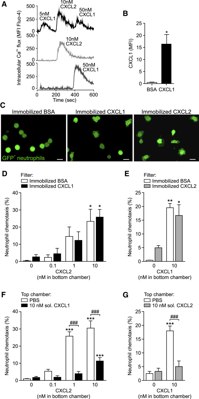
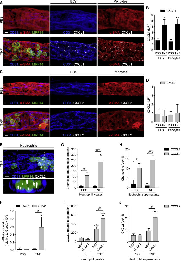
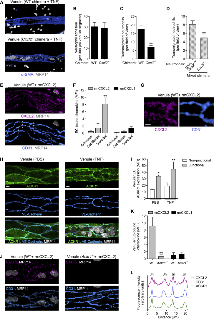
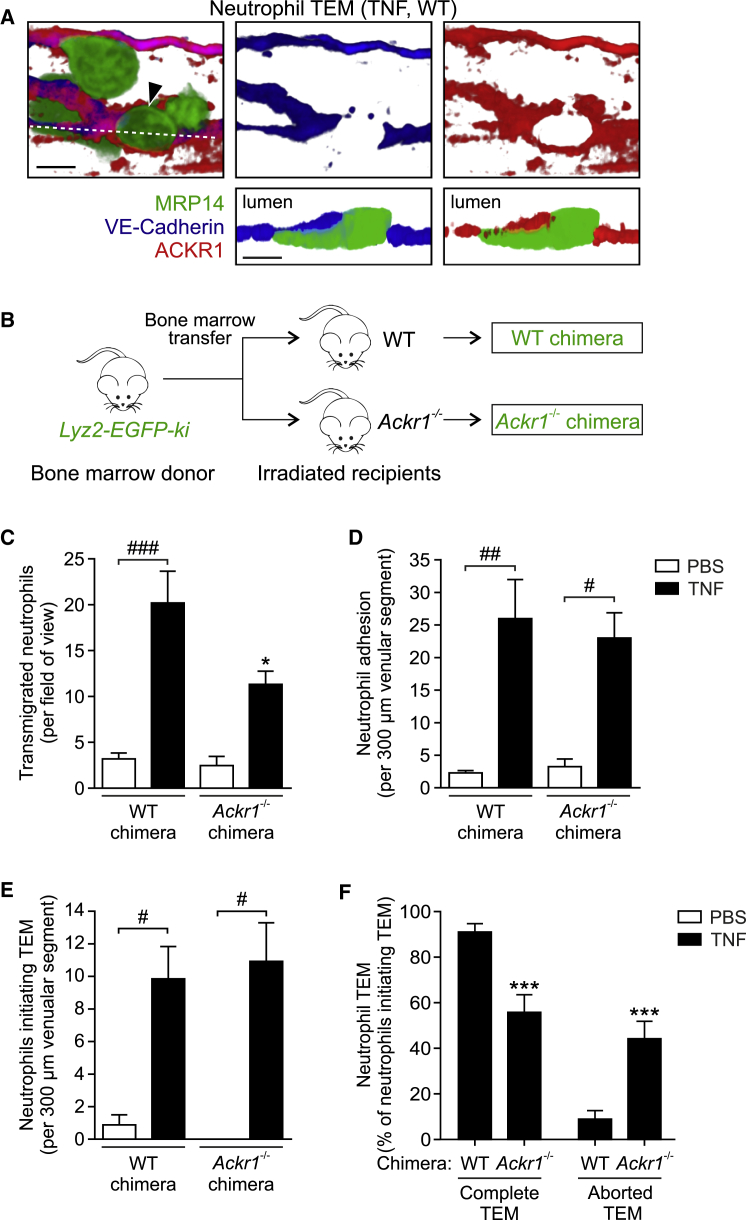
Neutrophils require directional cues to navigate through the complex structure of venular walls and into inflamed tissues. Here we applied confocal intravital microscopy to analyze neutrophil emigration in cytokine-stimulated mouse cremaster muscles. We identified differential and non-redundant roles for the chemokines CXCL1 and CXCL2, governed by their distinct cellular sources. CXCL1 was produced mainly by TNF-stimulated endothelial cells (ECs) and pericytes and supported luminal and sub-EC neutrophil crawling. Conversely, neutrophils were the main producers of CXCL2, and this chemokine was critical for correct breaching of endothelial junctions. This pro-migratory activity of CXCL2 depended on the atypical chemokine receptor 1 (ACKR1), which is enriched within endothelial junctions. Transmigrating neutrophils promoted a self-guided migration response through EC junctions, creating a junctional chemokine "depot" in the form of ACKR1-presented CXCL2 that enabled efficient unidirectional luminal-to-abluminal migration. Thus, CXCL1 and CXCL2 act in a sequential manner to guide neutrophils through venular walls as governed by their distinct cellular sources.

Keywords: ACKR1; CXCR2; chemokines; endothelium; extravasation; inflammation; neutrophils; pericytes.


Figures

None
Graphical abstract
Figure 1
Figure 1
TNF-Induced Neutrophil Migration Is Dependent on Both CXCL1 and CXCL2 WT mice pre-treated intrascrotally (i.s.) with control (ctr) or blocking mAbs or Cxcr2−/− mice were subjected to i.s. injections of PBS or TNF. The cremaster muscles were immunostained for MRP14 (neutrophils) and α-SMA (pericytes) and analyzed for neutrophil infiltration by confocal microscopy. (A) and (C) are representative images and (B), (D), and (E) show quantifications (n = 4–10 mice per group) from 5–6 independent experiments. Means ± SEM, ∗∗∗p < 0.001 as compared to TNF-treated ctrls and ###p < 0.001 as indicated. Scale bars, 30 μm. See also Figure S1.
Figure 2
Figure 2
TNF-Elicited CXCL1 and CXCL2 Support Distinct Phases of Neutrophil-EC Interactions Lyz2-EGFP-ki;Acta2-RFPcherry-Tg mice, subjected to in vivo CD31 labeling, were treated with ctr or blocking mAbs (i.v. 10 min prior to TNF in C and D and i.s. 100 min post TNF in E–H) and neutrophil responses in cremaster muscles injected locally with PBS or TNF quantified by confocal IVM. (A) Illustrative images of the employed IVM model (scale bar, 20 μm). (B) Scheme depicting neutrophil responses quantified in (C)–(H). (C and D) Quantification of neutrophil adhesion and intraluminal crawling (n = 5–6 mice per group, 23 independent experiments). (E) Time-lapse IVM images (Video S1) of a neutrophil TEM response in a TNF-stimulated tissue showing a neutrophil migrating from the lumen (0 min) through an EC junction (2–4 min) into the sub-EC space (6 min). Representative of 11 independent experiments; cross sections, top; luminal views, bottom; scale bars, 5 μm. (F) Quantifications of neutrophil TEM (n = 4–11 mice per group, 27 independent experiments). (G) IVM images (Video S2) of an aborted TEM response in a mouse treated with local TNF+anti-CXCL2 mAb. The images show a luminal neutrophil extending a protrusion through an EC junction (1 min), retracting the protrusion, and re-entering the circulation (5–8 min). Representative of 6 independent experiments; cross sections and luminal views; scale bars, 5 μm. (H) Quantification of aborted neutrophil TEM (n = 4–11 mice per group, 21 independent experiments). Means ± SEM, p < 0.05, ∗∗p < 0.01, ∗∗∗p < 0.001 as compared to TNF-treated ctrls and ##p < 0.01, ###p < 0.001 as indicated. See also Figure S2.
Figure 3
Figure 3
Neutrophil-Pericyte Interactions Are Selectively Mediated by Endogenous CXCL1 Lyz2-EGFP-ki;Acta2-RFPcherry-Tg mice, subjected to in vivo CD31 labeling, were stimulated locally with TNF and 100 min later i.s. injected with ctr or blocking mAbs, as indicated. (A) Neutrophil responses quantified in cremasteric venules by confocal IVM in (C)–(G). (B) Representative confocal IVM luminal and cross-sectional views depicting a neutrophil localized between TNF-stimulated ECs and pericytes 1 min post TEM. (C) Time-lapse IVM images (Video S3) showing a neutrophil crawling on pericytes (tracks, dashed lines) in a TNF-stimulated tissue (top) and the inhibition of this response in tissues treated with anti-CXCL1 mAb (bottom). Scale bars in (B) and (C), 10 μm. (D–G) Crawling profiles of neutrophils on pericytes (20 cells per group for clarity) (D) as normalized for their origin and associated quantifications of displacement (E), straightness index (displacement/track length) (F), and breaching of the pericyte layer (G). (H) Time-lapse confocal IVM images (Video S4) illustrating neutrophil reverse TEM in a tissue treated with TNF+anti-CXCL1 mAb (luminal and cross-sectional views; scale bars, 5 μm). (I) Quantifications of neutrophil reverse TEM. Images are representative of 5–10 independent experiments and quantifications (n = 5–10 mice per group) involve 20 independent experiments.
Figure 4
Figure 4
Neutrophils Can Respond to Sequential Exposure to CXCL1 and CXCL2 (A) Intracellular Ca2+ flux in neutrophils as induced by serial or single stimulations with chemokines representative of 4 independent experiments. (B) CXCL1 IF staining on Transwell filters coated with BSA or CXCL1 (n = 3) from 3 independent experiments. (C) Confocal images of GFP+ neutrophils seeded on BSA-, CXCL1-, or CXCL2-coated Transwell filters representative of 3 independent experiments. Scale bars, 10 μm. (D–G) Chemotaxis of neutrophils exposed to immobilized CXCL1 or CXCL2 (D and E) or soluble CXCL1 or CXCL2 (F and G) into bottom chambers containing indicated chemokines (1 hr at 37°C; n = 4) from 2–4 independent experiments. Means ± SEM, p < 0.05, ∗∗p < 0.01, ∗∗∗p < 0.001 as compared to corresponding responses without chemokines in bottom chambers and ###p < 0.001 as indicated. See also Figure S3.
Figure 5
Figure 5
CXCL1 and CXCL2 Are Differentially Expressed in TNF-Stimulated Tissues (A–E) WT mice were treated i.s. with PBS or TNF and cremaster muscles were IF stained for CXCL1 or CXCL2 and CD31 (ECs), α-SMA (pericytes), and MRP14 (neutrophils). Representative confocal images of venules showing CXCL1 (A) or CXCL2 (C) staining within EC and pericyte isosurface masks and quantifications of CXCL1 (B) and CXCL2 (D) in ECs or pericytes, in terms of MFIs (n = 4 mice per group) from 4 independent experiments. (E) Confocal abluminal and cross-sectional images (acquired along the dashed line and presented at 90° rotation) of a venule showing overall CXCL2 staining representative of 4 independent experiments. (F) Cxcl1 and Cxcl2 mRNA levels relative to Gapdh in circulating neutrophils 2–3 hr after i.s. PBS or TNF injection as determined by real-time PCR (n = 4–5 mice per group, 2 independent experiments). (G–J) Purified mouse bone marrow neutrophils were treated with PBS or TNF (1 nM, 1 hr) on uncoated (G, H), BSA-coated, or CXCL1-coated wells (I, J). CXCL1 and/or CXCL2 levels in lysates (G and I) and supernatants (H and J) as quantified by ELISA (n = 4–12) from 2–4 independent experiments. Means ± SEM, p < 0.05, ∗∗p < 0.01, ∗∗∗p < 0.001 as compared to corresponding PBS-treated ctrls and #p < 0.05, ##p < 0.01, ###p < 0.001, as indicated. Scale bars, 5 μm. See also Figure S4.
Figure 6
Figure 6
CXCL2 Mediates Neutrophil TEM in a Cell-Autonomous Manner and Is Retained at EC Junctions via the Atypical Chemokine Receptor ACKR1 (A–C) Representative confocal images of venules in TNF-stimulated cremaster muscles of control (WT) or leukocyte CXCL2-deficient (Cxcl2−/−) chimeras stained for α-SMA and MRP14 (A) and associated quantifications of neutrophil adhesion and extravasation (B, C; n = 4 mice per group) from 4 independent experiments). Scale bars, 30 μm. (D) Numbers of extravasated GFP-Cxcl2wt/wt and Cxcl2−/− neutrophils in TNF-stimulated cremaster muscles of mixed chimeras, as normalized for neutrophil numbers in the blood (n = 4 mice, 3 independent experiments). (E–G) RmCXCL2 or rmCXCL1 was injected i.s. into WT mice and cremaster muscles were stained for CXCL2 or CXCL1, CD31, and MRP14 and analyzed by confocal microscopy. Images (E; representative of 4 independent experiments; scale bar, 5 μm), quantified localization of rmCXCL2 or rmCXCL1 within the microcirculation (F, n = 3–8 mice per group, 8 independent experiments), and enlarged images of the boxed region in (E) (G; scale bar, 2 μm). (H and I) Cremaster muscles were stimulated with PBS or TNF and IF stained for ACKR1, VE-Cadherin, and MRP14. Confocal images of venules (H; scale bars, 5 μm) and quantification of ACKR1 localization (I, n = 4 mice per group, 4 independent experiments). (J–L) IF localization of rmCXCL2 and rmCXCL1 in cremasteric venules in relation to ACKR1 expression. Representative confocal images of tissues from WT and Ackr1−/− mice injected with rmCXCL2 (J; scale bars, 5 μm) and quantifications of endothelial rmCXCL2 and rmCXCL1 binding (K, n = 4–5 mice per group, 3 independent experiments). (L) CXCL2, ACKR1, and CD31 IF intensities along the dashed line in (E) cutting across 4 EC junctions (Jn; representative of 4 independent experiments). Means ± SEM, p < 0.05, ∗∗p < 0.01 (as compared to arterioles or capillaries in F and non-junctional ACKR1 expression in I). See also Figure S5.
Figure 7
Figure 7
Endothelial ACKR1 Facilitates Decisive Luminal-to-Abluminal TEM (A) Confocal images showing a neutrophil (arrow) migrating through an EC junction in a TNF-stimulated cremasteric venule of a WT mouse IF stained for MRP14, VE-Cadherin, and ACKR1. Luminal (top) and cross-sectional images (bottom) along dashed line; representative of 3 independent experiments; scale bars, 5 μm. (B) Scheme illustrating the generation of chimeric mice exhibiting Lyz2-EGFP-ki hematopoietic cells (GFP+ neutrophils) and WT or Ackr1−/− non-hematopoietic cells. (C–F) Quantification of neutrophil extravasation (C), adhesion to ECs (D), initiating TEM (E), and undergoing complete or aborted TEM (F) in cremaster muscles of WT and Ackr1−/− chimeras after local PBS or TNF injection (n = 3–8 mice per group, 26 independent experiments) as analyzed by confocal IVM. Means ± SEM, p < 0.05, ∗∗∗p < 0.001 relative to WT chimeras and #p < 0.05, ##p < 0.01, ###p < 0.001 as indicated. See also Figure S6.

Comment in

References

    1. Armstrong D.A., Major J.A., Chudyk A., Hamilton T.A. Neutrophil chemoattractant genes KC and MIP-2 are expressed in different cell populations at sites of surgical injury. J. Leukoc. Biol. 2004;75:641–648. - PubMed
    1. Cacalano G., Lee J., Kikly K., Ryan A.M., Pitts-Meek S., Hultgren B., Wood W.I., Moore M.W. Neutrophil and B cell expansion in mice that lack the murine IL-8 receptor homolog. Science. 1994;265:682–684. - PubMed
    1. Chou R.C., Kim N.D., Sadik C.D., Seung E., Lan Y., Byrne M.H., Haribabu B., Iwakura Y., Luster A.D. Lipid-cytokine-chemokine cascade drives neutrophil recruitment in a murine model of inflammatory arthritis. Immunity. 2010;33:266–278. - PMC - PubMed
    1. Colom B., Bodkin J.V., Beyrau M., Woodfin A., Ody C., Rourke C., Chavakis T., Brohi K., Imhof B.A., Nourshargh S. Leukotriene B4-neutrophil elastase axis drives neutrophil reverse transendothelial cell migration in vivo. Immunity. 2015;42:1075–1086. - PMC - PubMed
    1. Dawson T.C., Lentsch A.B., Wang Z., Cowhig J.E., Rot A., Maeda N., Peiper S.C. Exaggerated response to endotoxin in mice lacking the Duffy antigen/receptor for chemokines (DARC) Blood. 2000;96:1681–1684. - PubMed

Publication types

MeSH terms