

A20 rescues hepatocytes from apoptosis through the NF-κB signaling pathway in rats with acute liver failure
- PMID: 30446523
- PMCID: PMC6328859
- DOI: 10.1042/BSR20180316
A20 rescues hepatocytes from apoptosis through the NF-κB signaling pathway in rats with acute liver failure
Retraction in
-
Retraction: A20 rescues hepatocytes from apoptosis through the NF-κB signaling pathway in rats with acute liver failure.Biosci Rep. 2021 Oct 29;41(10):BSR-20180316_RET. doi: 10.1042/BSR-2018-0316_RET. Biosci Rep. 2021. PMID: 34647576 Free PMC article. No abstract available.
Abstract
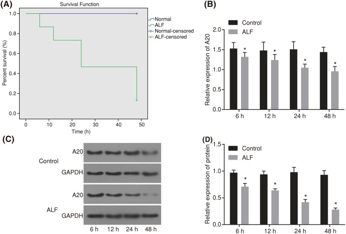
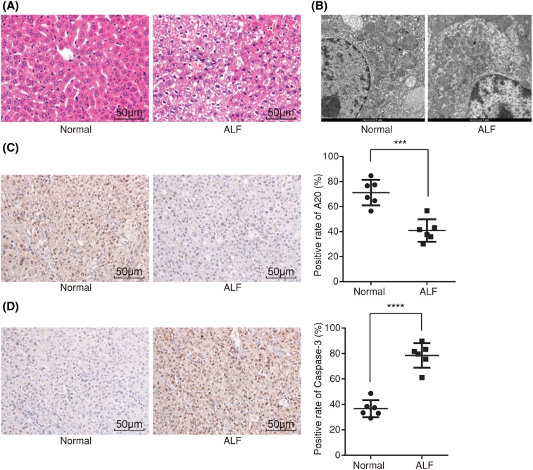
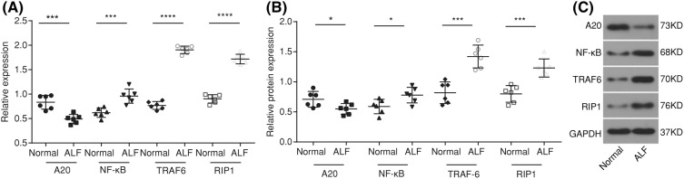
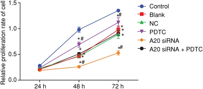
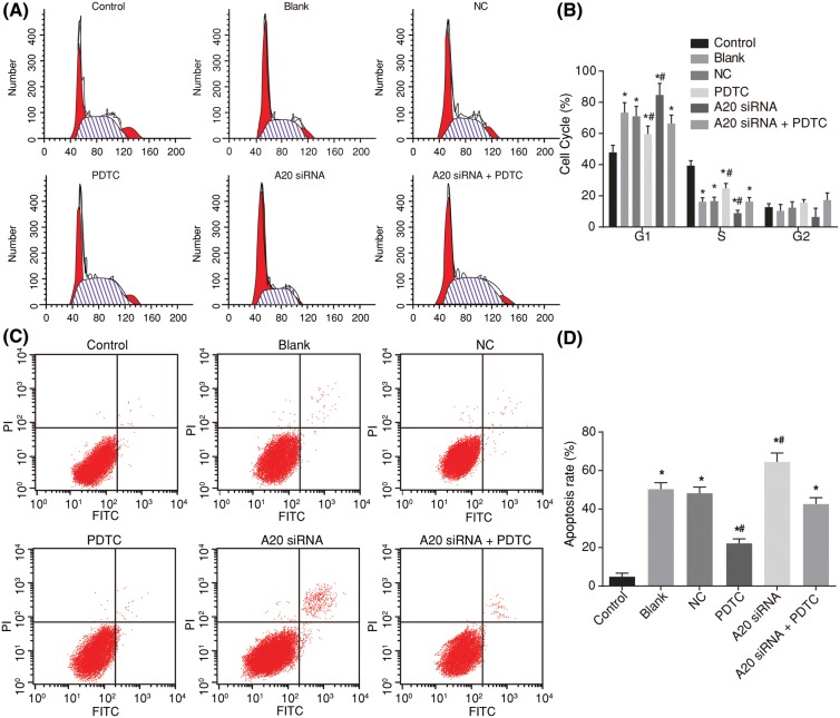
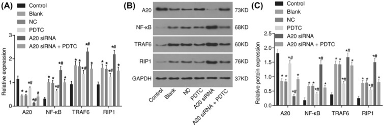
Background: Acute liver failure (ALF) is a disease of acute derangements in the hepatic synthetic function with defects involving innate immune responses, which was reported to be negatively regulated by tumor necrosis factor α-induced protein 3 (A20). Herein, the present study was conducted to investigate the effects the A20 protein on the proliferation and apoptosis of hepatocytes through the nuclear factor (NF)-κB signaling pathway in the rat models simulating ALF.Methods: Male Wistar rats were used to simulate ALF in the model rats. Next, the positive expression of A20 and Caspase-3 proteins was measured in liver tissues. Rat hepatocytes were separated and subjected to pyrrolidine dithiocarbamate (PDTC, inhibitor of NF-κB pathway) or A20 siRNA. Additionally, both mRNA and protein levels of A20, NF-κB, tumor necrosis factor (TNF) receptor-associated factor 6 (TRAF6), and receptor-interacting protein 1 (RIP1) were determined. Finally, we detected the hepatocyte proliferation, cell cycle entry, and apoptosis.Results: ALF rats displayed a lower positive expression of A20 protein and a higher expression of Caspase-3 protein. Furthermore, A20 was down-regulated, while NF-κB, TRAF6, and RIP1 were all up-regulated in ALF rats. Notably, A20 inhibited activation of NF-κB signaling pathway. The blockade of NF-κB signaling pathway enhanced proliferation and cell cycle progression of hepatocytes, whereas inhibited apoptosis of hepatocytes. On the contrary, A20 siRNA reversed the above situation.Conclusion: A20 inhibits apoptosis of hepatocytes and promotes the proliferation through the NF-κB signaling pathway in ALF rats, potentially providing new insight into the treatment of ALF.
Keywords: A20; Acute liver failure; Hepatocyte; NF-κB signaling pathway; Proliferation; Tumor necrosis factor α-induced protein 3.
© 2018 The Author(s).
Conflict of interest statement
The authors declare that there are no competing interests associated with the manuscript.
Figures
Similar articles
-
Lipoxin A4 exerts protective effects against experimental acute liver failure by inhibiting the NF-κB pathway.Int J Mol Med. 2016 Mar;37(3):773-80. doi: 10.3892/ijmm.2016.2483. Epub 2016 Feb 5. Int J Mol Med. 2016. PMID: 26865215
-
A20 overexpression inhibits lipopolysaccharide-induced NF-κB activation, TRAF6 and CD40 expression in rat peritoneal mesothelial cells.Int J Mol Sci. 2014 Apr 17;15(4):6592-608. doi: 10.3390/ijms15046592. Int J Mol Sci. 2014. PMID: 24747594 Free PMC article.
-
The zinc finger protein A20 inhibits TNF-induced NF-kappaB-dependent gene expression by interfering with an RIP- or TRAF2-mediated transactivation signal and directly binds to a novel NF-kappaB-inhibiting protein ABIN.J Cell Biol. 1999 Jun 28;145(7):1471-82. doi: 10.1083/jcb.145.7.1471. J Cell Biol. 1999. PMID: 10385526 Free PMC article.
-
ABINs: A20 binding inhibitors of NF-kappa B and apoptosis signaling.Biochem Pharmacol. 2009 Jul 15;78(2):105-14. doi: 10.1016/j.bcp.2009.02.009. Epub 2009 Feb 27. Biochem Pharmacol. 2009. PMID: 19464428 Review.
-
The biology of A20-binding inhibitors of NF-kappaB activation (ABINs).Adv Exp Med Biol. 2014;809:13-31. doi: 10.1007/978-1-4939-0398-6_2. Adv Exp Med Biol. 2014. PMID: 25302363 Review.
Cited by
-
Regulation of apoptosis by ubiquitination in liver cancer.Am J Cancer Res. 2023 Oct 15;13(10):4832-4871. eCollection 2023. Am J Cancer Res. 2023. PMID: 37970337 Free PMC article. Review.
-
Chinese Herb Jiedu Huayu Granules Inhibiting Immune and Inflammatory Response of Rats with Acute Liver Failure by Regulating the NF-κB Signaling Pathway.Biomed Res Int. 2022 May 11;2022:4479885. doi: 10.1155/2022/4479885. eCollection 2022. Biomed Res Int. 2022. Retraction in: Biomed Res Int. 2023 Dec 29;2023:9853907. doi: 10.1155/2023/9853907. PMID: 35601154 Free PMC article. Retracted.
-
A potential link between plasma short-chain fatty acids, TNF-α level and disease progression in non-alcoholic fatty liver disease: A retrospective study.Exp Ther Med. 2022 Jul 28;24(3):598. doi: 10.3892/etm.2022.11536. eCollection 2022 Sep. Exp Ther Med. 2022. PMID: 35949337 Free PMC article.
-
Electroacupuncture and Parecoxib Reduce Inflammatory Injury in a Primary Dysmenorrhea Rat Model: Investigating the Role of the COX-2/NF-κB/NLRP3 Pathway.J Pain Res. 2025 Jul 15;18:3573-3592. doi: 10.2147/JPR.S512243. eCollection 2025. J Pain Res. 2025. PMID: 40687333 Free PMC article.
-
A20 Establishes Negative Feedback With TRAF6/NF-κB and Attenuates Early Brain Injury After Experimental Subarachnoid Hemorrhage.Front Immunol. 2021 Jul 26;12:623256. doi: 10.3389/fimmu.2021.623256. eCollection 2021. Front Immunol. 2021. PMID: 34381441 Free PMC article.
References
Publication types
MeSH terms
Substances
LinkOut - more resources
Full Text Sources
Research Materials
Miscellaneous