. 2019 Jan 2;39(1):63-77.
doi: 10.1523/JNEUROSCI.2095-18.2018. Epub 2018 Nov 16.

Glial Sulfatides and Neuronal Complex Gangliosides Are Functionally Interdependent in Maintaining Myelinating Axon Integrity

Affiliations

Glial Sulfatides and Neuronal Complex Gangliosides Are Functionally Interdependent in Maintaining Myelinating Axon Integrity

Rhona McGonigal et al. J Neurosci. .

Abstract

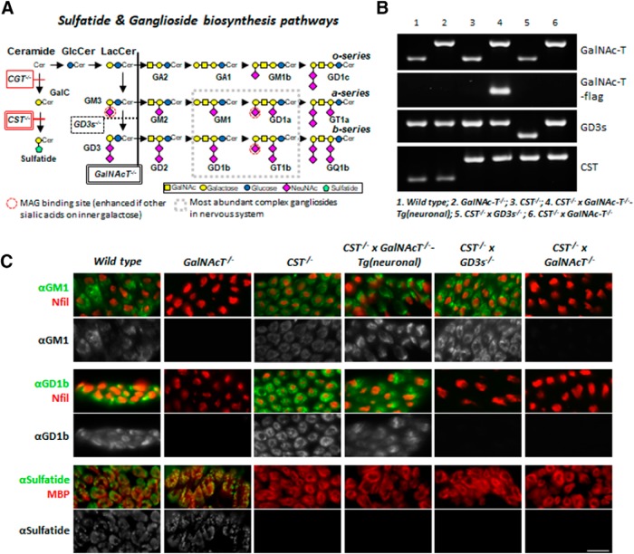
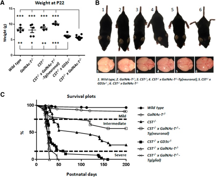
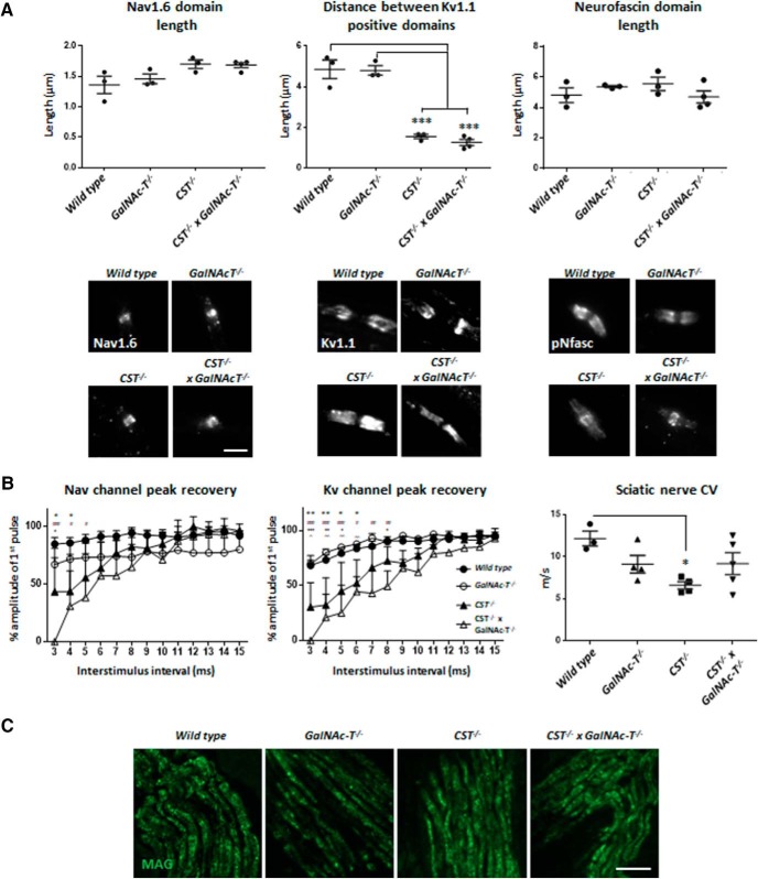
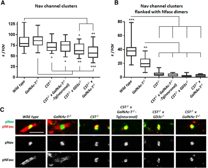
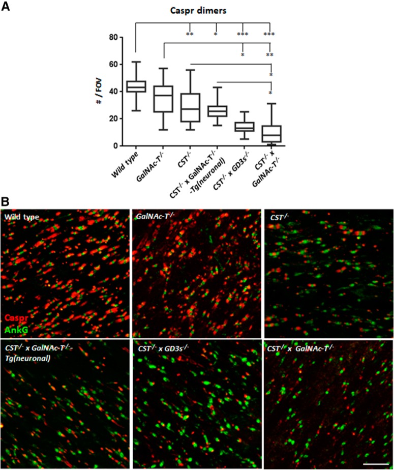
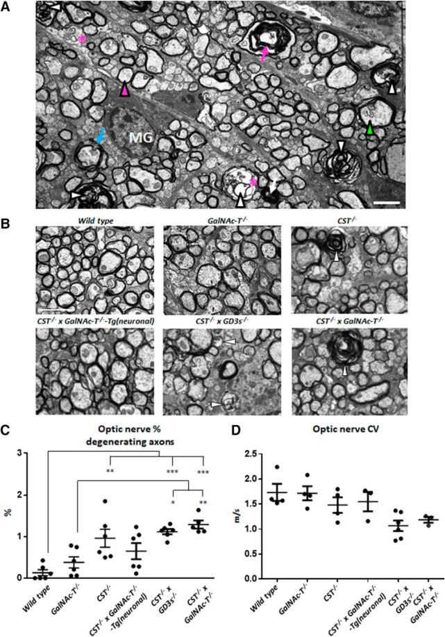
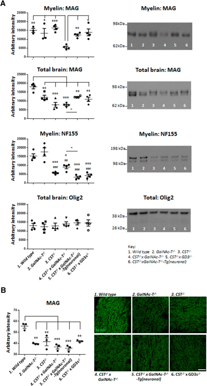
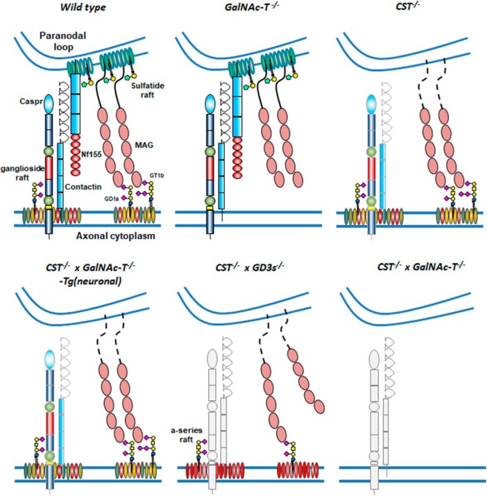
Sulfatides and gangliosides are raft-associated glycolipids essential for maintaining myelinated nerve integrity. Mice deficient in sulfatide (cerebroside sulfotransferase knock-out, CST-/-) or complex gangliosides (β-1,4-N-acetylegalactosaminyltransferase1 knock-out, GalNAc-T-/-) display prominent disorganization of proteins at the node of Ranvier (NoR) in early life and age-dependent neurodegeneration. Loss of neuronal rather than glial complex gangliosides underpins the GalNAc-T-/- phenotype, as shown by neuron- or glial-specific rescue, whereas sulfatide is principally expressed and functional in glial membranes. The similarities in NoR phenotype of CST-/-, GalNAc-T-/-, and axo-glial protein-deficient mice suggests that these glycolipids stabilize membrane proteins including neurofascin155 (NF155) and myelin-associated glycoprotein (MAG) at axo-glial junctions. To assess the functional interactions between sulfatide and gangliosides, CST-/- and GalNAc-T-/- genotypes were interbred. CST-/-× GalNAc-T-/- mice develop normally to postnatal day 10 (P10), but all die between P20 and P25, coinciding with peak myelination. Ultrastructural, immunohistological, and biochemical analysis of either sex revealed widespread axonal degeneration and disruption to the axo-glial junction at the NoR. In addition to sulfatide-dependent loss of NF155, CST-/- × GalNAc-T-/- mice exhibited a major reduction in MAG protein levels in CNS myelin compared with WT and single-lipid-deficient mice. The CST-/- × GalNAc-T-/- phenotype was fully restored to that of CST-/- mice by neuron-specific expression of complex gangliosides, but not by their glial-specific expression nor by the global expression of a-series gangliosides. These data indicate that sulfatide and complex b-series gangliosides on the glial and neuronal membranes, respectively, act in concert to promote NF155 and MAG in maintaining the stable axo-glial interactions essential for normal nerve function.SIGNIFICANCE STATEMENT Sulfatides and complex gangliosides are membrane glycolipids with important roles in maintaining nervous system integrity. Node of Ranvier maintenance in particular requires stable compartmentalization of multiple membrane proteins. The axo-glial adhesion molecules neurofascin155 (NF155) and myelin-associated glycoprotein (MAG) require membrane microdomains containing either sulfatides or complex gangliosides to localize and function effectively. The cooperative roles of these microdomains and associated proteins are unknown. Here, we show vital interdependent roles for sulfatides and complex gangliosides because double (but not single) deficiency causes a rapidly lethal phenotype at an early age. These findings suggest that sulfatides and complex gangliosides on opposing axo-glial membranes are responsible for essential tethering of the axo-glial junction proteins NF155 and MAG, which interact to maintain the nodal complex.

Keywords: MAG; NF155; axo–glial integrity; ganglioside; node of Ranvier; sulfatide.


Figures

Figure 1.
Figure 1.
Generation of lipid-deficient transgenic mouse lines and confirmation by PCR and immunostaining. Six mouse lines were used and generated: WT, GalNAc-T−/−, CST−/−, CST−/− × GalNAc-T−/−-Tg(neuronal), CST−/− × GD3s−/−, and CST−/− × GalNAc-T−/−. A, Sulfatide and ganglioside biosynthesis pathways (associated gene knock-outs are indicated in boxes). Ceramide is the precursor to sulfatide and gangliosides. The CST enzyme is necessary for the synthesis of sulfatide from GalC. The GalNAc-T enzyme is necessary for generation of complex gangliosides and the GD3s enzyme for specific production of b-series complex gangliosides. Constructs were generated to drive GalNAc-T expression in neurons of GalNAc-T−/− × CST−/− mice to produce the GalNAc-T−/−-Tg(neuronal) mouse line. Location of MAG binding site to terminal 2,3 sialic acid is indicated by a red circle. B, PCR results confirming the presence or absence of GalNAc-T, GD3s, and CST expression in mouse lines. Large bands represent the disrupted GalNAc-T and CST gene with insert; the smaller band represents the disruption of the GD3s gene. The flag identifies the reintroduction of the GalNAc-T gene into the neurons. C, Expression of a- and b-series gangliosides and sulfatide was confirmed by staining peripheral nerves with anti-GM1, anti-GD1b, and anti-sulfatide monoclonal antibodies (green), respectively. Axons and myelin were identified with neurofilament or MBP (red), respectively. Scale bar, 10 μm.
Figure 2.
Figure 2.
Altering the expression of gangliosides and sulfatide in novel transgenic mouse lines effects survival and phenotype. A, Weight of double-null (n = 10) and CST−/− × GD3s−/− (n = 6) mice is normal during development, but declines from P15–P25 and is significantly reduced compared with other genotypes at P22. Statistical differences among genotypes were determined by one-way ANOVA followed by Tukey's post hoc tests to compare multiple comparisons, indicated on the graphs as follows: *p < 0.05; **p < 0.01; ***p < 0.001. B, CST−/− × GalNAc-T−/−, and CST−/− × GD3s−/− mice display a reduction in stature, hindlimb leg splaying, a hunched appearance, and tremor at P22. Gross brain anatomy does not differ between genotypes. C, Survival plots demonstrate that incrementally diminishing ganglioside and sulfatide expression corresponds to a reduction in life expectancy of up to 200 d. WT (n = 7) and GalNAc-T−/− (n = 10) mice have a normal life expectancy. In the absence of sulfatide with normal ganglioside expression, life expectancy is more than halved in CST−/− mice (n = 18). Interestingly, in the absence of sulfatide and complex gangliosides with the reintroduction of complex gangliosides into neurons alone (n = 6), life expectancy is improved, with 60% of mice surviving to 200 d and beyond. Mice with no sulfatide and a-series gangliosides expressed globally (n = 38) can survive up to 20 weeks. Double ganglioside and sulfatide knock-out mice (n = 29) have the worst phenotype, never surviving past 4 weeks and dying at P21–P25. Reintroduction of gangliosides into glia (n = 12) does not improve survival.
Figure 3.
Figure 3.
The additional loss of complex gangliosides on a sulfatide-null background does not augment disorganization at the NoR or electrophysiological function in the peripheral nervous system. A, There is modest Nav1.6 channel cluster domain lengthening in both CST−/− and CST−/− × GalNAc-T−/− teased SNs that does not reach significance. Representative images from teased SNs immunostained for Nav1.6 show that Nav channel cluster appearance was similar in every genotype. Disturbance of the paranode is indicated by invasion of Kv1.1 channels into the paranode, shown by a decrease in the gap between positive domains. The gap between Kv1.1-positive domains is significantly and comparably decreased in both CST−/− and CST−/− × GalNAc-T−/− mice compared with WT and GalNAc-T−/− NoRs. Representative images show that Kv1.1 formed two distinct domains of immunostaining at the juxtaparanodes in WT and GalNAcT−/− mice. Conversely, mice lacking sulfatide expression had Kv1.1 staining at the paranodes, suggesting disruption to the axo–glial junction. The length of pNFasc-immunostained domains does not significantly differ among genotypes. However, representative images show that labeling greatly differs: WT and GalNAc-T−/− NoRs have pNFasc staining that is most intense at the paranodes, forming two clear dimers (“normal”), whereas both CST−/− and CST−/− × GalNAc-T−/− mice have intense staining at the nodal gap (presumed NF186) with weaker paranodal (presumed NF155) staining (“abnormal”), which indicates a disturbance in paranodal adhesion (WT, n = 3; GalNAc-T−/−, n = 3; CST−/−, n = 3; and CST−/− × GalNAc-T−/−, n = 4). Scale bar, 5 μm. B, Perineural recordings of Nav and Kv channel currents from intercostal nerves showing an increase in recovery time for both peaks following paired pulse stimulation in both CST−/− and CST−/− × GalNAc-T−/− mice compared with WT and GalNAc-T−/− mice. The CST−/− and CST−/− × GalNAc-T−/− mice were not significantly different from each other at any ISI. Graphs display the means and ±SEM for each genotype (WT n = 5, GalNAc-T−/− n = 2, CST−/− n = 3, CST−/− × GalNAc-T−/− n = 2) and statistical analysis performed (two-way ANOVA; #WT vs CST−/− × GalNAc-T−/− mice; *WT vs CST−/−; +GalNAc-T−/− vs CST−/− × GalNAc-T−/−; ∧GalNAc-T−/− vs CST−/− × GalNAc-T−/−). SN conduction velocity decreased in CST−/− (n = 4), GalNAc-T−/− (n = 4), and CST−/− × GalNAc-T−/− (n = 5) mice compared with WT (n = 3), but only reached significance in CST−/− nerves. C, Peripheral nerve MAG immunostaining was comparable among genotypes. Scale bar, 20 μm. Graphs display the means and ±SEM for each genotype and statistical differences among genotypes were determined by one-way ANOVA followed by Tukey's post hoc tests to compare multiple comparisons, indicated on the graphs as follows: *p < 0.05; **p < 0.01; ***p < 0.001.
Figure 4.
Figure 4.
Loss of both sulfatide and complex gangliosides results in a modest reduction in CNS Nav channel cluster number and sulfatide deficiency reduces NF155 presence at paranodal loops. A, The number of Nav channel clusters significantly decreases in glycolipid deficient mouse OpNs (n = 3–4/genotype). Reintroducing a- and b-series gangliosides into neurons rescues this feature. B, Nav channel clusters flanked by normal paranodal NF dimers are significantly reduced in number when sulfatide is not expressed (n = 2–3/genotype). Box-and-whisker plots are used to display the spread of all data points collected from each animal and means were calculated per genotype for statistical analysis (one-way ANOVA followed by Tukey's post hoc tests to compare multiple comparisons, indicated on the graphs as follows: *p < 0.05; **p < 0.01; ***p < 0.001). C, Representative images of OpN sections from each genotype double-immunostained for pan-Nav (pNav) antibody (green) and pNFasc antibody (red). Na channel clusters were observed in every genotype. pNFasc immunostaining formed a long band crossing the node and paranodes in WT and GalNAc-T−/− mice, suggesting labeling of the NF186 and NF155 isoforms, respectively. All of the genotypes lacking sulfatide expression had pNFasc staining restricted to the NoR and colocalizing only with pNav staining, suggesting the presence of only the NF186 isoform of NF. Scale bar, 5 μm.
Figure 5.
Figure 5.
Paranodal Caspr dimer immunostaining in CNS tissue is progressively reduced with increasing glycolipid deficiency. A, CST−/− × GalNAc-T−/− and CST−/− × GD3s−/− mice have significantly fewer Caspr dimers per FOV compared with WT and GalNAc-T−/− mice and are not significantly different from each other (n = 2–5/genotype). Caspr dimer number is improved to levels within GalNAc-T−/− mice range, but not WT, in CST−/− and CST−/− × GalNAc-T−/−-Tg(neuronal) mice, which both display significantly more Caspr dimers than CST−/− × GalNAc-T−/− mice. Box-and-whisker plots are used to display the spread of all data points collected from each animal and means were calculated per genotype for statistical analysis (one-way ANOVA followed by Tukey's post hoc tests to compare multiple comparisons, indicated on the graphs as follows: *p < 0.05; **p < 0.01; ***p < 0.001). B, Representative images of OpN sections from each genotype double-immunostained for Caspr (red) and the nodal marker AnkyrinG (green) showing the reduction in Caspr dimer number flanking ankyrin G clusters with diminishing glycolipid expression. Scale bar, 10 μm.
Figure 6.
Figure 6.
Glycolipid deficiency causes pathological changes and compromises CNS axon survival and function. A, Representative ultrastructural features observed in CST−/− × GD3s−/− and CST−/− × GalNAc-T−/− mice. Shown are normal myelinated axon (magenta arrowhead), degenerating axons (white arrowheads), dark condensed degenerating axon (white arrow), vacuoles within axons (*), empty myelin sheath (magenta arrow), redundant myelin (blue arrow), abnormal cytoskeleton (green arrowhead), and microglia (MG). B, Electron micrographs depicting the differences among the genotypes. White arrowheads indicate degenerating axons. Myelin is similar among mouse lines. C, Degenerating axon number increases to a significant level compared with WT in OpNs from CST−/−, CST−/− × GD3s−/−, and CST−/− × GalNAc-T−/− mice. Reintroducing a- and b-series gangliosides into neurons rescues this pathology. D, Reduction in conduction velocity was observed in OpNs with diminishing glycolipid content. This did not reach significance between genotypes. One-way ANOVA followed by Tukey's post hoc tests to compare multiple comparisons, indicated on the graphs as follows: *p < 0.05; **p < 0.01; ***p < 0.001. Scale bar, 2 μm.
Figure 7.
Figure 7.
MAG and NF155 expression in the myelin fraction is altered by glycolipid deficiency. Note the new order of genotypes compared to other figures. A, In the myelin fraction from P22 brain homogenates, MAG is significantly reduced in CST−/− × GalNAc-T−/− mice compared with all genotypes, which do not significantly differ from one another. All genotypes have a significant reduction in MAG expression in whole-brain homogenate compared with WT. Myelin NF155 was significantly reduced in all genotypes compared with WT (*) and GalNAc-T−/− (#), which had similar levels. ∧Significant difference between CST−/− × GalNAc-T−/− and CST−/− × GalNAc-T−/−-Tg(neuronal) mice. Olig2 is unchanged by altered glycolipid expression. Representative blots to the right of corresponding graphs show protein intensity per genotype, as indicated by the corresponding number in the key below. B, P22 OpN MAG immunostaining is significantly reduced in all genotypes compared with WT nerves, which do not differ significantly from one another. Representative images show MAG immunostaining per genotype. Means were calculated per genotype for statistical analysis (one-way ANOVA followed by Tukey's post hoc tests to compare multiple comparisons, indicated on the graphs as follows: *p < 0.05; **p < 0.01; ***p < 0.001). Scale bar, 50 μm. ##p < 0.01, ###p < 0.001, p < 0.05.
Figure 8.
Figure 8.
Schematic depicting the glycolipid rafts and their associated paranodal proteins Caspr, NF155, and MAG under WT conditions and in our transgenic mouse lines. GD1a and GT1b are represented in the ganglioside rafts because they are the major ligands of MAG; however, in reality, the rafts would contain all complex gangliosides. Normally, GD1a and GT1b in rafts will tether MAG, but, in their absence, MAG does not make the axo–glial connection. If NF155 is present, then this protein can partner with Caspr/contactin to make an axo–glial junction. However, when sulfatide is absent, NF155 is also lost from the paranode. In the absence of NF155, Caspr is also diminished, especially with loss of complex ganglioside rafts. Under conditions of both ganglioside and sulfatide deficiency, we propose an absence of the structurally supporting proteins MAG and NF155 that results in the loss of a functionally competent axo–glial junction.

Comment in

Similar articles

Cited by

References

    1. Barrie JA, Montague P, Karim S, Kirkham D, Nave KA, Anderson TJ, Griffiths IR, McLaughlin M (2010) Modulation of rumpshaker phenotype with wild-type PLP/DM20 suggests several pathogenic mechanisms. J Neurosci Res 88:2135–2145. 10.1002/jnr.22379 - DOI - PubMed
    1. Bartsch U, Kirchhoff F, Schachner M (1989) Immunohistological localization of the adhesion molecules L1, N-CAM, and MAG in the developing and adult optic nerve of mice. J Comp Neurol 284:451–462. 10.1002/cne.902840310 - DOI - PubMed
    1. Bhat MA, Rios JC, Lu Y, Garcia-Fresco GP, Ching W, St Martin M, Li J, Einheber S, Chesler M, Rosenbluth J, Salzer JL, Bellen HJ (2001) Axon-glia interactions and the domain organization of myelinated axons requires neurexin IV/Caspr/Paranodin. Neuron 30:369–383. 10.1016/S0896-6273(01)00294-X - DOI - PubMed
    1. Boffey J, Odaka M, Nicoll D, Wagner ER, Townson K, Bowes T, Conner J, Furukawa K, Willison HJ (2005) Characterisation of the immunoglobulin variable region gene usage encoding the murine anti-ganglioside antibody repertoire. J Neuroimmunol 165:92–103. 10.1016/j.jneuroim.2005.04.011 - DOI - PubMed
    1. Bowes T, Wagner ER, Boffey J, Nicholl D, Cochrane L, Benboubetra M, Conner J, Furukawa K, Furukawa K, Willison HJ (2002) Tolerance to self-gangliosides is the major factor restricting the antibody response to lipopolysaccharide core oligosaccharides in campylobacter jejuni strains associated with guillain-barre syndrome. Infect Immun 70:5008–5018. 10.1128/IAI.70.9.5008-5018.2002 - DOI - PMC - PubMed

Publication types

MeSH terms

LinkOut - more resources