

Sialic Acid-Dependent Inhibition of T Cells by Exosomal Ganglioside GD3 in Ovarian Tumor Microenvironments
- PMID: 30446565
- PMCID: PMC6289713
- DOI: 10.4049/jimmunol.1801041
Sialic Acid-Dependent Inhibition of T Cells by Exosomal Ganglioside GD3 in Ovarian Tumor Microenvironments
Abstract
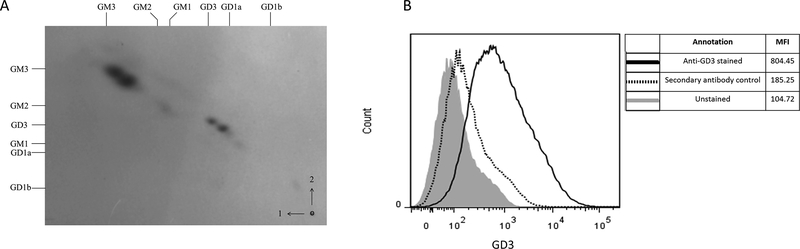
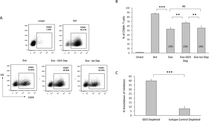
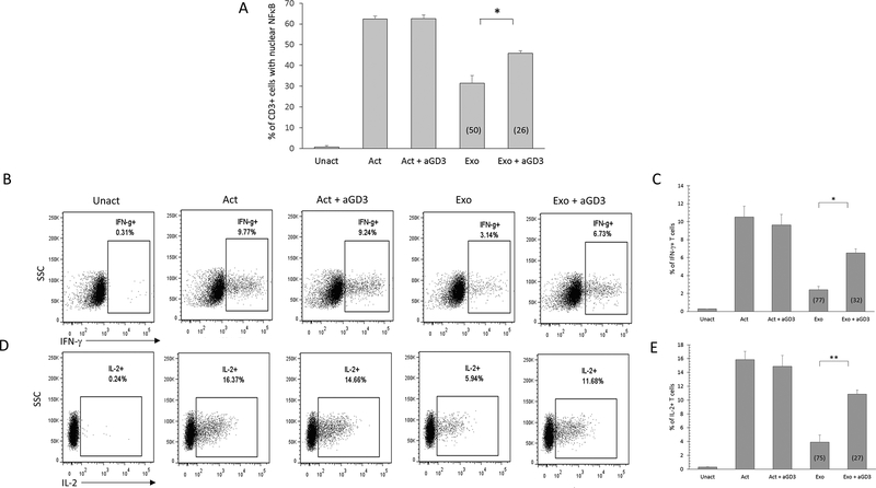
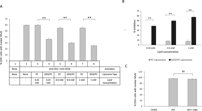
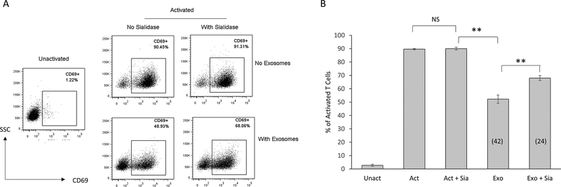
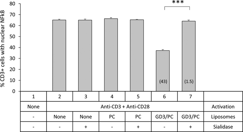
The tumor microenvironment is rendered immunosuppressive by a variety of cellular and acellular factors that represent potential cancer therapeutic targets. Although exosomes isolated from ovarian tumor ascites fluids have been previously reported to induce a rapid and reversible T cell arrest, the factors present on or within exosomes that contribute to immunosuppression have not been fully defined. In this study, we establish that GD3, a ganglioside expressed on the surface of exosomes isolated from human ovarian tumor ascites fluids, is causally linked to the functional arrest of T cells activated through their TCR. This arrest is inhibited by Ab blockade of exosomal GD3 or by the removal of GD3+ exosomes. Empty liposomes expressing GD3 on the surface also inhibit the activation of T cells, establishing that GD3 contributes to the functional arrest of T cells independent of factors present in exosomes. Finally, we demonstrate that the GD3-mediated arrest of the TCR activation is dependent upon sialic acid groups, because their enzymatic removal from exosomes or liposomes results in a loss of inhibitory capacity. Collectively, these data define GD3 as a potential immunotherapeutic target.
Copyright © 2018 by The American Association of Immunologists, Inc.
Conflict of interest statement
Figures
Similar articles
-
Exosomes Represent an Immune Suppressive T Cell Checkpoint in Human Chronic Inflammatory Microenvironments.Immunol Invest. 2020 Oct;49(7):726-743. doi: 10.1080/08820139.2020.1748047. Epub 2020 Apr 17. Immunol Invest. 2020. PMID: 32299258 Free PMC article.
-
Human ovarian tumor ascites fluids rapidly and reversibly inhibit T cell receptor-induced NF-κB and NFAT signaling in tumor-associated T cells.Cancer Immun. 2013 Jul 15;13:14. Print 2013. Cancer Immun. 2013. PMID: 23882159 Free PMC article.
-
Novel phosphatidylserine-binding molecule enhances antitumor T-cell responses by targeting immunosuppressive exosomes in human tumor microenvironments.J Immunother Cancer. 2021 Oct;9(10):e003148. doi: 10.1136/jitc-2021-003148. J Immunother Cancer. 2021. PMID: 34599030 Free PMC article.
-
Tumor-Associated Exosomes: A Potential Therapeutic Target for Restoring Anti-Tumor T Cell Responses in Human Tumor Microenvironments.Cells. 2021 Nov 13;10(11):3155. doi: 10.3390/cells10113155. Cells. 2021. PMID: 34831378 Free PMC article. Review.
-
Aiming for the Sweet Spot: Glyco-Immune Checkpoints and γδ T Cells in Targeted Immunotherapy.Front Immunol. 2020 Sep 29;11:564499. doi: 10.3389/fimmu.2020.564499. eCollection 2020. Front Immunol. 2020. PMID: 33133075 Free PMC article. Review.
Cited by
-
Plasma Exosomes of Patients with Breast and Ovarian Tumors Contain an Inactive 20S Proteasome.Molecules. 2021 Nov 18;26(22):6965. doi: 10.3390/molecules26226965. Molecules. 2021. PMID: 34834058 Free PMC article.
-
Extracellular vesicles in ovarian cancer chemoresistance, metastasis, and immune evasion.Cell Death Dis. 2022 Jan 18;13(1):64. doi: 10.1038/s41419-022-04510-8. Cell Death Dis. 2022. PMID: 35042862 Free PMC article. Review.
-
Prominosomes - a particular class of extracellular vesicles containing prominin-1/CD133?J Nanobiotechnology. 2025 Jan 29;23(1):61. doi: 10.1186/s12951-025-03102-w. J Nanobiotechnology. 2025. PMID: 39881297 Free PMC article. Review.
-
Neurological aspects of SARS-CoV-2 infection: lipoproteins and exosomes as Trojan horses.Trends Endocrinol Metab. 2022 Aug;33(8):554-568. doi: 10.1016/j.tem.2022.04.011. Epub 2022 May 2. Trends Endocrinol Metab. 2022. PMID: 35613979 Free PMC article. Review.
-
The Role and Clinical Interest of Extracellular Vesicles in Pregnancy and Ovarian Cancer.Biomedicines. 2021 Sep 18;9(9):1257. doi: 10.3390/biomedicines9091257. Biomedicines. 2021. PMID: 34572444 Free PMC article. Review.
References
-
- Santin AD, Bellone S, Ravaggi A, Pecorelli S, Cannon MJ, and Parham GP 2000. Induction of ovarian tumor-specific CD8+ cytotoxic T lymphocytes by acid-eluted peptide-pulsed autologous dendritic cells. Obstet Gynecol 96: 422–430. - PubMed
-
- Ramakrishna V, Ross MM, Petersson M, Gatlin CC, Lyons CE, Miller CL, Myers HE, McDaniel M, Karns LR, Kiessling R, Parmiani G, and Flyer DC 2003. Naturally occurring peptides associated with HLA-A2 in ovarian cancer cell lines identified by mass spectrometry are targets of HLA-A2-restricted cytotoxic T cells. Int Immunol 15: 751–763. - PubMed
-
- Zhang L, Conejo-Garcia JR, Katsaros D, Gimotty PA, Massobrio M, Regnani G, Makrigiannakis A, Gray H, Schlienger K, Liebman MN, Rubin SC, and Coukos G 2003. Intratumoral T cells, recurrence, and survival in epithelial ovarian cancer. N Engl J Med 348: 203–213. - PubMed
-
- Yokota SJ, Facciponte JG, Kelleher RJ Jr., Shultz LD, Loyall JL, Parsons RR, Odunsi K, Frelinger JG, Lord EM, Gerber SA, Balu-Iyer SV, and Bankert RB 2013. Changes in ovarian tumor cell number, tumor vasculature, and T cell function monitored in vivo using a novel xenograft model. Cancer Immun 13: 11. - PMC - PubMed
-
- Moon EK, Wang LC, Dolfi DV, Wilson CB, Ranganathan R, Sun J, Kapoor V, Scholler J, Pure E, Milone MC, June CH, Riley JL, Wherry EJ, and Albelda SM 2014. Multifactorial T-cell hypofunction that is reversible can limit the efficacy of chimeric antigen receptor-transduced human T cells in solid tumors. Clin Cancer Res 20: 4262–4273. - PMC - PubMed
Publication types
MeSH terms
Substances
Grants and funding
LinkOut - more resources
Full Text Sources
Other Literature Sources
Medical
Research Materials
