. 2019 Feb;368(2):169-178.
doi: 10.1124/jpet.118.252650. Epub 2018 Nov 16.

Nicotine Modulates Growth Factors and MicroRNA to Promote Inflammatory and Fibrotic Processes

Affiliations

Nicotine Modulates Growth Factors and MicroRNA to Promote Inflammatory and Fibrotic Processes

Afshin Ebrahimpour et al. J Pharmacol Exp Ther. 2019 Feb.

Abstract

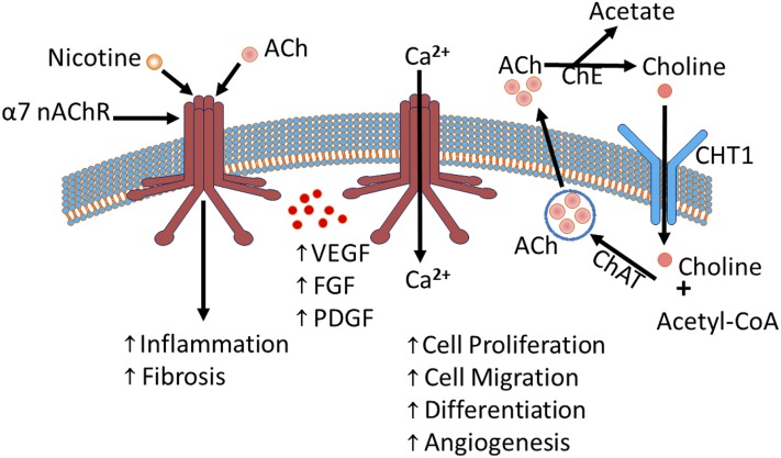
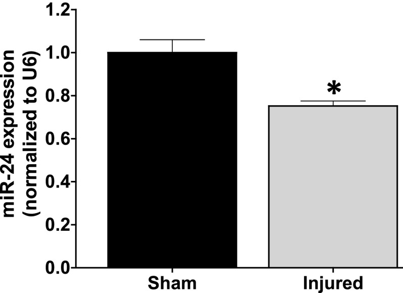
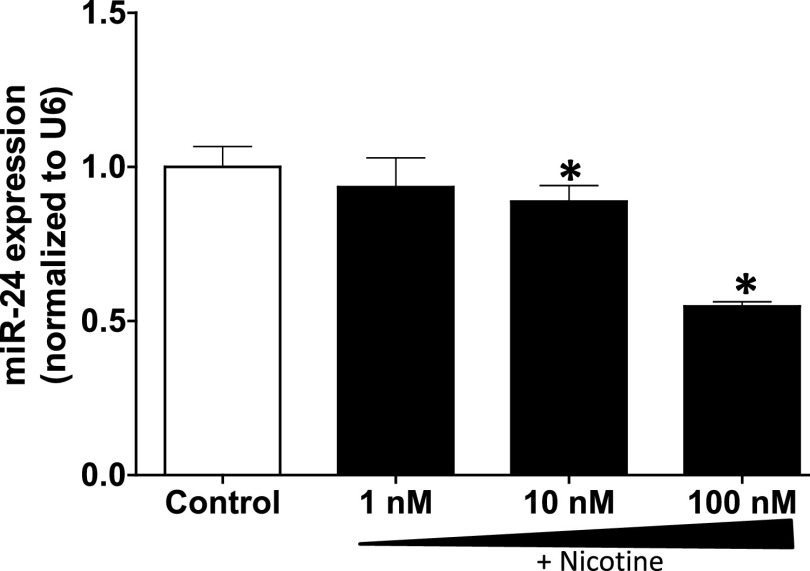
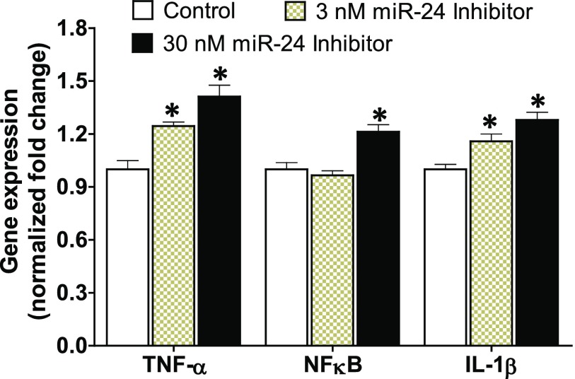
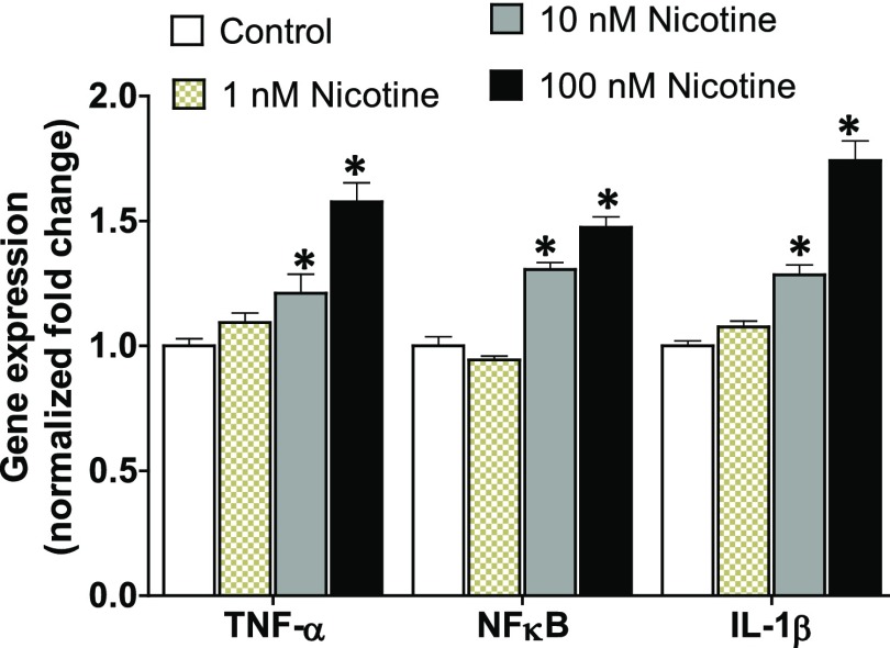
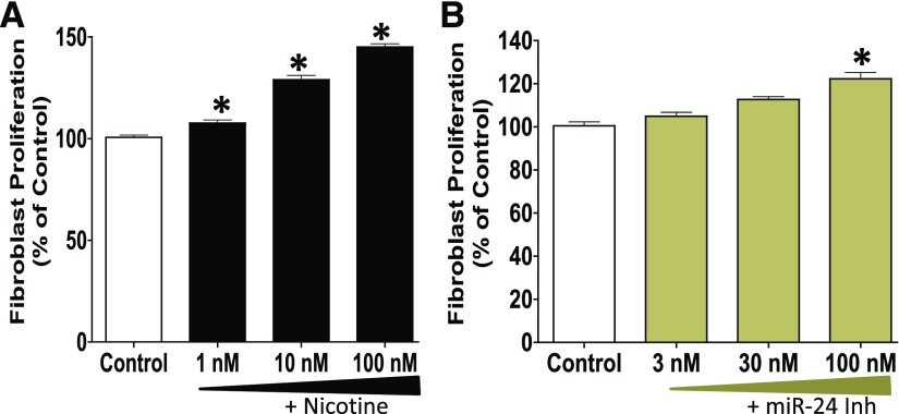
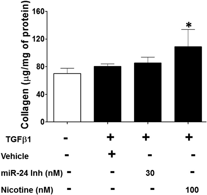
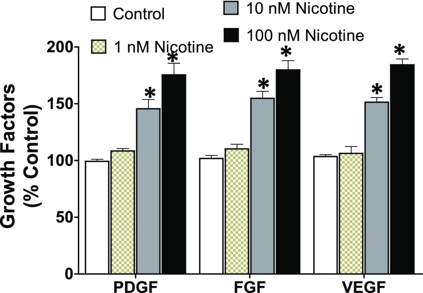
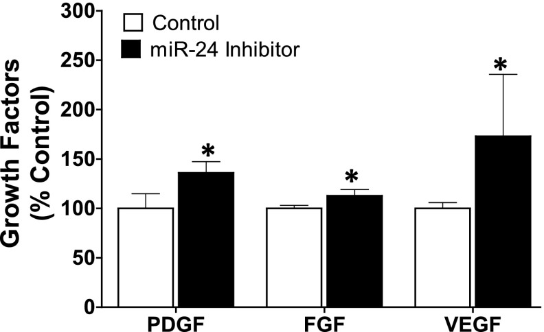
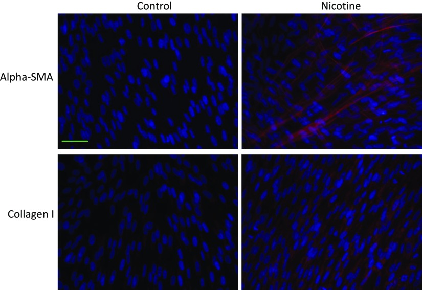
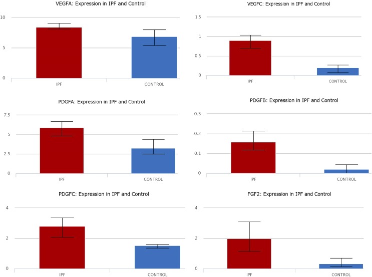
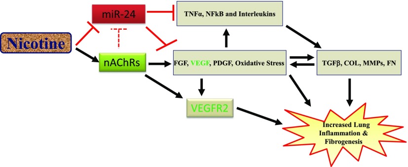
Idiopathic pulmonary fibrosis (IPF) is a fatal disease that destroys the structure and function of the lungs. Risk factors include advanced age and genetic predisposition. However, tobacco use is the chief modifiable risk factor. The prevalence of tobacco use in IPF reaches up to 80%. Although tobacco smoke contains over 5000 chemicals, nicotine is a major component. Nicotine is a bioactive molecule that acts upon nicotinic acetylcholine receptors expressed on neuronal and non-neuronal cells including endothelial cells. Accordingly, it has a pleiotropic effect on cell proliferation and angiogenesis. The angiogenic effect is partly mediated by stimulation of growth factors including fibroblast, platelet-derived, and vascular endothelial growth factors. Nintedanib, a Food and Drug Administration-approved drug for IPF, works by inhibiting receptors for these growth factors, suggesting a pathobiologic role of the growth factors in IPF and a potential mechanism by which tobacco use may exacerbate the disease process; additionally, nicotine downregulates anti-inflammatory microRNAs (miRs) in lung cells. Here, we profiled the expression of miRs in lung tissues explanted from a lung injury model and examined the effect of nicotine on one of the identified miRs (miR-24) and its downstream targets. Our data show that miR-24 is downregulated during lung injury and is suppressed by nicotine. We also found that nicotine upregulates the expression of inflammatory cytokines targeted by miR-24. Finally, nicotine stimulated growth factors, fibroblast proliferation, collagen release, and expression of myofibroblast markers. Taken together, nicotine, alone or as a component of tobacco smoke, may accelerate the disease process in IPF through stimulation of growth factors and downregulation of anti-inflammatory miRs.


Figures

Fig. 1.
Fig. 1.
The nicotinic acetylcholine machinery: ACh or nAChRs, including the homomeric α7 nAChR promote growth factors (VEGF, FGF, and PDGF) and influence several physiologic processes including cell proliferation, migration, differentiation, and angiogenesis, as well as pathologic conditions, including inflammation and fibrosis. Permeability of the α7 nAChR to cations (e.g., Ca2+) is shown. In addition, the degradation of ACh into choline, the internalization of choline and its recycling to form ACh molecules is shown. Ca2+, calcium; ChAT, choline acetyltransferase; ChE, choline esterase; CHT1, choline transporter.
Fig. 2.
Fig. 2.
miR-24 expression is reduced in lungs explanted from animal model of bleomycin-induced lung injury. Lung injury was induced in F344 Fischer rats through a single intratracheal administration of bleomycin at 4 mg/kg body weight. Animals were euthanized 28-days post-bleomycin injury and the right lung lobes were used for miR expression studies. Data are mean ± S.E.M. from duplicate experiments (n = 3). The noncoding small nuclear RNA U6 was used as the loading control and to normalize the expression of miR-24 in the samples. *P < 0.05 vs. sham.
Fig. 3.
Fig. 3.
Nicotine dose dependently downregulates the expression of miR-24 in lung epithelial cells. Primary human lung epithelial cells were treated with nicotine (1–100 nM) or control (PBS) for 24 hours prior to isolating total RNA containing miRs. The expression of miR-24 was amplified by reverse transcription polymerase chain reaction. Data are mean ± S.E.M. from duplicate experiments (n = 3). U6 was used as the loading control and to normalize the expression of miR-24 in the samples. *P < 0.05 vs. control.
Fig. 4.
Fig. 4.
Inhibition of miR-24 in human lung epithelial cells upregulates the expression of inflammatory cytokines: TNF-α, NFκB, and IL-1β. Primary human lung epithelial cells were treated with miR-24 inhibitor (3–30 nM) or control for 24 hours prior to isolating total RNA. The expression of proinflammatory genes was amplified by reverse transcription polymerase chain reaction. The housekeeping gene β-actin was used as the internal control and data were normalized to the control group. Data are mean ± S.E.M. from duplicate experiments (n = 4). *P < 0.05 vs. control.
Fig. 5.
Fig. 5.
Nicotine induces the expression of proinflammatory cytokines in lung epithelial cells. Human lung epithelial cells were treated with nicotine (1–100 nM) or control for 24 hours. Total RNA was isolated and the expression of TNF-α, NFκB, and IL-1β was profiled. β-actin was used as the internal control and data were normalized to the control group. Data are mean ± S.E.M. from duplicate experiments. *P < 0.05 vs. control (n = 4).
Fig. 6.
Fig. 6.
The proliferation of lung fibroblasts is increased upon treatment with: (A) nicotine (1–100 nM) or (B) miR-24 inhibitor (3–100 nM). Synchronized primary human lung fibroblasts (seeding density = 3 × 10³) were treated with nicotine, miR-24 inhibitor, or control for 24 hours and allowed to proliferate in the presence of BrdU for an additional 22 hours. Cell proliferation was assessed using BrdU assay. Data are mean ± S.E.M. from duplicate experiments (n = 3) and are expressed as the percentage of proliferation in the control groups. *P < 0.05 vs. control; miR-24 Inh denotes miR-24 inhibitor.
Fig. 7.
Fig. 7.
Nicotine enhances soluble collagen release by lung fibroblasts. Primary human lung fibroblasts were seeded at 6 × 10⁴ cells/well in six-well plates. Synchronized cells were treated with recombinant human TGFβ1 (10 ng/ml) in the presence of nicotine (100 nM), miR-24 inhibitor (30 nM), or vehicle for 24 hours. Acid-soluble collagen content in each well was determined in the conditioned media using Sircol collagen assay. The amount of collagen in each well was estimated from a standard curve and the collagen content in each sample was normalized to total cellular protein. Data are expressed as microgram collagen per milligram of protein. Data are mean ± S.E.M. from duplicate experiments (n = 3). *P < 0.05 compared with no TGFβ control.
Fig. 8.
Fig. 8.
Nicotine increases the expression of growth factors PDGF, FGF, and VEGF in lung endothelial cells. Primary human lung endothelial cells were seeded on 75 cm2 flasks until 80% confluency, and then treated with nicotine (1–100 nM) or control (PBS) for 24 hours. The concentration of the growth factors in the conditioned media was measured by ELISA (optical density: 450 nm readout) and calculated from the standard curve. Data were normalized to total cellular protein and are expressed as mean ± S.E.M. from duplicate experiments (n = 3). *P < 0.05 vs. control.
Fig. 9.
Fig. 9.
Inhibition of miR-24 increases the expression of growth factors PDGF, FGF, and VEGF in lung endothelial cells. Primary human lung endothelial cells were seeded on 75 cm2 flasks until 80% confluency, and then treated with miR-24 inhibitor (100 nM) or control miR for 24 hours. The intracellular growth factor concentration was measured by ELISA (optical density: 450 nm readout) and calculated from the standard curve. Data were normalized to total cellular protein and are expressed as mean ± S.E.M. from duplicate experiments (n = 3). *P < 0.05 vs. control.
Fig. 10.
Fig. 10.
Immunofluorescence stain showing increased expression of the myofibroblast markers α-SMA and collagen I upon chronic exposure of lung fibroblasts to nicotine. Primary human lung fibroblasts were seeded in six-well plates and treated with nicotine (100 nM) daily for 5 days. The cells were fixed, permeabilized, and stained for the myofibroblast markers: α-SMA (1:400; Abcam), and collagen I (1:500; Abcam) (n = 3). Goat anti-rabbit (1:500; ThermoFisher) Alexa Fluor 594 secondary antibodies were used and are shown as red stain. The nuclei were stained with 4′,6-diamidino-2-phenylindole and are shown as blue stain. Objective original magnification, 40×; Scale bar, 50 µm (applies to all the images in this figure).
Fig. 11.
Fig. 11.
The gene expression of VEGF (isoforms A and C); PDGF (isoforms A, B, and C); and FGF-2 is increased in lung epithelial cells isolated from IPF patients compared with controls. Data are curated from RNA sequencing analysis from the lung gene expression analysis web portal comparing single cells isolated from IPF patients and control lungs.
Fig. 12.
Fig. 12.
Schematic showing the potential mechanism of accelerated lung inflammation and fibrosis by nicotine. As shown, nicotine internalizes through the endogenous nAChRs to promote growth factors (VEGF, FGF, and PDGF) and induce oxidative stress, which in turn stimulates profibrotic molecules (TGFβ, collagen, fibronectin, and metalloproteinases). In addition, we propose that nicotine suppresses anti-inflammatory microRNA such as miR-24 to derepress the expression of inflammatory cytokines including TNFα, NFκB, and interleukins, which are known to interact with mitogenic growth factors and profibrotic molecules to promote aberrant lung remodeling.

Similar articles

Cited by

References

    1. Akram KM, Lomas NJ, Forsyth NR, Spiteri MA. (2014) Alveolar epithelial cells in idiopathic pulmonary fibrosis display upregulation of TRAIL, DR4 and DR5 expression with simultaneous preferential over-expression of pro-apoptotic marker p53. Int J Clin Exp Pathol 7:552–564. - PMC - PubMed
    1. Allen JT, Spiteri MA. (2002) Growth factors in idiopathic pulmonary fibrosis: relative roles. Respir Res 3:13. - PMC - PubMed
    1. Antoniades HN, Bravo MA, Avila RE, Galanopoulos T, Neville-Golden J, Maxwell M, Selman M. (1990) Platelet-derived growth factor in idiopathic pulmonary fibrosis. J Clin Invest 86:1055–1064. - PMC - PubMed
    1. Atamas SP. (2017) Vascular endothelial growth factor in idiopathic pulmonary fibrosis. An imbalancing act. Am J Respir Crit Care Med 196:409–411. - PubMed
    1. Barr J, Sharma CS, Sarkar S, Wise K, Dong L, Periyakaruppan A, Ramesh GT. (2007) Nicotine induces oxidative stress and activates nuclear transcription factor kappa B in rat mesencephalic cells. Mol Cell Biochem 297:93–99. - PMC - PubMed

Publication types

MeSH terms