. 2019 Feb;18(2):235-244.
doi: 10.1158/1535-7163.MCT-18-0768. Epub 2018 Nov 16.

Induction of MNK Kinase-dependent eIF4E Phosphorylation by Inhibitors Targeting BET Proteins Limits Efficacy of BET Inhibitors

Affiliations

Induction of MNK Kinase-dependent eIF4E Phosphorylation by Inhibitors Targeting BET Proteins Limits Efficacy of BET Inhibitors

Thao N D Pham et al. Mol Cancer Ther. 2019 Feb.

Abstract

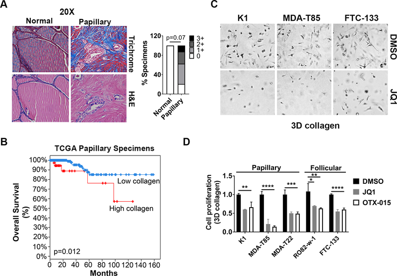
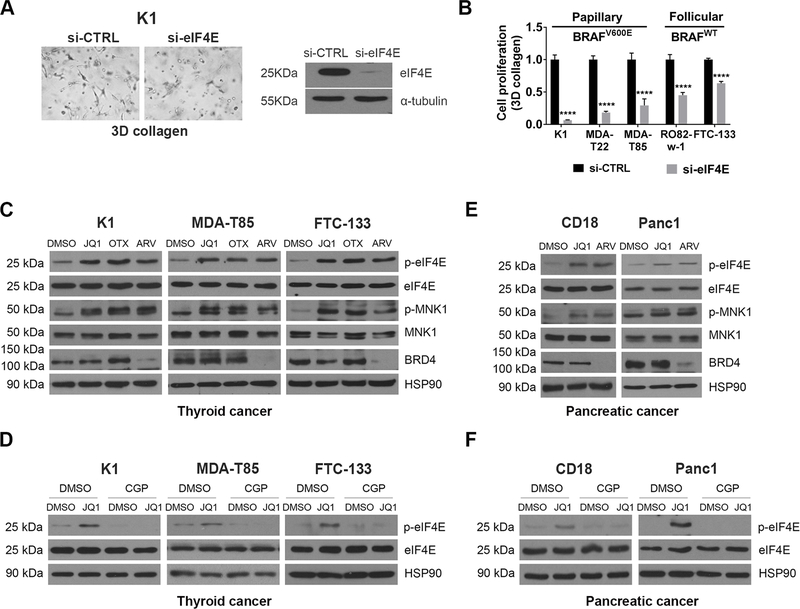
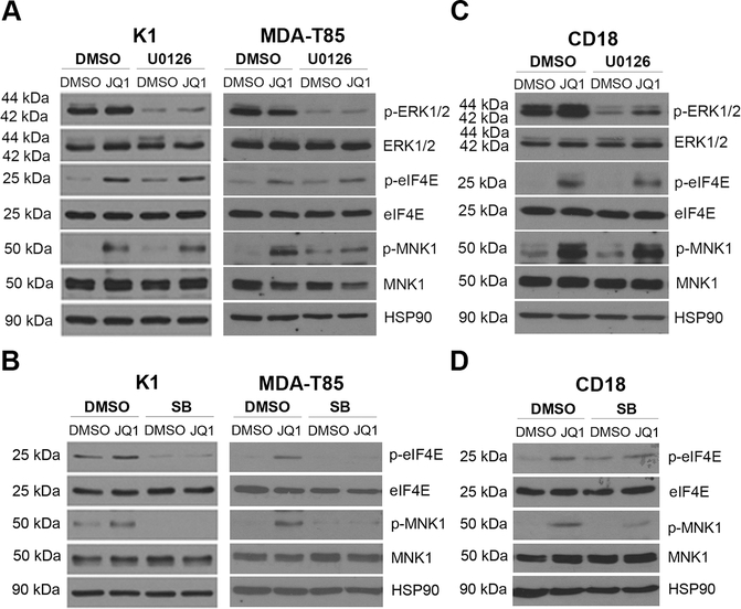
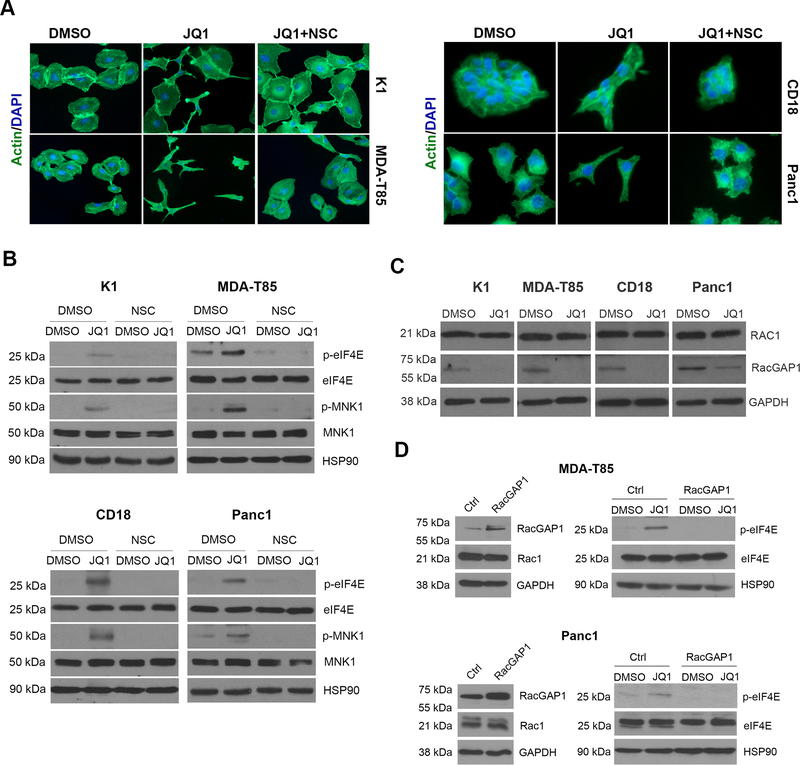
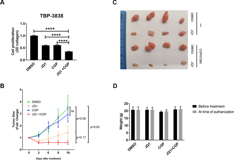
BET inhibitors (BETi), which target transcription of key oncogenic genes, are currently being evaluated in early-phase clinical trials. However, because BETis show limited single-agent activity, there is increasing interest in identifying signaling pathways to enhance the efficacy of BETis. Here, we demonstrate increased MNK kinase-dependent eIF4E phosphorylation following treatment with BETis, indicating activation of a prosurvival feedback mechanism in response to BETis. BET PROTACs, which promote degradation of BET proteins, also induced eIF4E phosphorylation in cancer cells. Mechanistically, we show that the effect of BETis on MNK-eIF4E phosphorylation was mediated by p38 MAPKs. We also show that BETis suppressed RacGAP1 to induce Rac signaling-mediated eIF4E phosphorylation. Significantly, MNK inhibitors and MNK1/2 knockdown enhanced the efficacy of BETis in suppressing proliferation of cancer cells in vitro and in a syngeneic mouse model. Together, these results demonstrate a novel prosurvival feedback signaling induced by BETis, providing a mechanistic rationale for combination therapy with BET and MNK inhibitors for synergistic inhibition of cancer cells.


Conflict of interest statement

Disclosure of Potential Conflicts of Interest

The authors declare no potential conflicts of interest.

Figures

Figure 1:
Figure 1:. BET inhibitors (BETis) decrease growth of cancer cells in 3D collagen.
A. Human thyroid tissue microarray (TMA) containing 24 papillary thyroid specimens and 8 adjacent normal tissue samples was trichrome stained, and the relative staining was graded as low (0 or 1+) or high (2+ or 3+). Fisher’s exact test was used to calculate p value. B. cBioportal analysis of Kaplan-Meier survival curve of papillary thyroid cancer patients in the TCGA database with or without relative overexpression of fibrillar collagen. C and D. Thyroid cancer cells growing in 3D collagen were treated with the BETis JQ1 (1 μM) and OTX-015 (1 μM) for 72 hours. The cells were examined by phase microscopy (C), and the effect on proliferation was determined by WST-1 assay (D). *, p<0.05; **, p<0.01; ***, p<0.001; ****, p<0.0001. The results are representative of at least three independent experiments.
Figure 2:
Figure 2:. BETis and BET PROTACs induce eIF4E phosphorylation.
A. Thyroid cancer cells were transfected with siRNA targeting eIF4E for 48 hours, and then cultured in 3D collagen for additional 48 hours. The cells were examined by phase microscopy. Knockdown efficiency was determined by Western blotting. B. Thyroid cancer cells with or without BRAFV600E mutation were transfected with siRNA targeting eIF4E for 48 hours and then cultured in 3D collagen for additional 48 hours. The effect on proliferation was determined by WST-1 assay. ****, p<0.0001. C. Thyroid cancer cells were treated with BETis JQ1 (1 μM) and OTX-015 (OTX, 1 μM) or the BET PROTAC ARV-825 (ARV, 1 μM) for 24 hours. The effect on eIF4E and MNK1 phosphorylation and expression of eIF4E, MNK1, BRD4 and HSP90 (loading control) was determined by Western blotting. D. Thyroid cancer cells were pre-treated with CGP57380 (CGP, 10 μM) for 30 min, followed by treatment of JQ1 (1 μM) for 24 hours. The effect on eIF4E phosphorylation and expression of eIF4E and HSP90 was determined by Western blotting. E. CD18 and Panc1 pancreatic cancer cells were treated with the BET inhibitor JQ1 (1 μM) or the BET PROTAC ARV-825 (ARV, 1 μM) for 24 hours. The effect on eIF4E and MNK1 phosphorylation and expression of eIF4E, MNK1, BRD4 and HSP90 (loading control) was determined by Western blotting. F. Pancreatic cancer cells were pre-treated with CGP57380 (CGP, 10 μM) for 30 min and then treated with JQ1 (1 μM) and for 24 hours. The effect on eIF4E phosphorylation and expression of eIF4E and HSP90 was determined by Western blotting. The results are representative of at least three independent experiments.
Figure 3:
Figure 3:. p38 MAPKs, but not MEK/ERK signaling, mediates BET inhibitor-induced eIF4E phosphorylation.
A and C. Thyroid and pancreatic cancer cells were pre-treated with the MEK1/2 inhibitor U0126 (5 μM) for 30 min and then treated with JQ1 (1 μM) and for 24 hours. The effect on ERK1/2, MNK1 and eIF4E phosphorylation and on the expression of ERK1/2, MNK1 and eIF4E expression was determined by Western blotting, using HSP90 as loading control. B and D. Thyroid and pancreatic cancer cells were pre-treated with the p38 MAPK inhibitor SB202190 (SB, 5 μM) for 30 min and then treated with JQ1 (1 μM) and for 24 hours. The effect on MNK1 and eIF4E phosphorylation and on the expression of MNK1 and eIF4E expression was determined by Western blotting, using HSP90 as loading control. The results are representative of at least three independent experiments.
Figure 4:
Figure 4:. BETis induce Rac-mediated cytoskeletal changes and Rac-mediated eIF4E phosphorylation.
A. Cancer cells growing on glass coverslips were treated with DMSO or JQ1 (1 μM) and co-treated with the Rac inhibitor NSC23766 (NSC, 50 μM) for 24 hours. The cells were then processed for phalloidin staining and the nuclei counterstained with DAPI (40x). B. Cancer cells were pre-treated with the Rac inhibitor NSC23766 (NSC, 50 μM) and then treated with JQ1 (1 μM) for 24 hours. The effect on eIF4E and MNK1 phosphorylation was determined by Western blotting. C. Cancer cells were treated with DMSO or JQ1 (1 μM) for 24 hours and the effect on Rac1 and RacGAP1 was determined by Western blotting. D. Cancer cells transfected with control (Ctrl) vector or vector expressing RacGAP1 were treated with DMSO or JQ1 (1 μM) for 24 hours. The effect on eIF4E phosphorylation and on the expression of RacGAP1, Rac1 and eIF4E expression was determined by western blotting, using GAPDH and HSP90 as loading controls. The results are representative of at least three independent experiments.
Figure 5:
Figure 5:. MNK inhibitors and MNK1/2 siRNA potentiate the effects of BETis at suppressing proliferation.
A and B. Cancer cells growing in 3D collagen were treated with JQ1 (1 μM) and co-treated with CGP57380 (CGP, 2 μM) for 72 hours and the effect on cell proliferation was determined using the WST-1 assay. ***, p<0.001; ****, p<0.0001 relative to the combination treatment group. C-F. Cancer cells were transfected with control siRNA (siCTRL) or combination of siRNAs against MNK1 and MNK2 (siMNK1/2) for 48 hours. The cells were then embedded in 3D collagen and treated with JQ1 (1 μM) for 48 hours. The effect on MNK1 and MNK2 knockdown was determined by Western blotting and/or by qRT-PCR. The effect on eIF4E and MNK1 phosphorylation was determined by Western blotting using HSP90 as loading control (C-D). The effect on cell proliferation was determined using the WST-1 assay (E, F). ****, p<0.0001 relative to siCTRL-transfected, DMSO-treated samples. The results are representative of three independent experiments.
Figure 6:
Figure 6:. MNK inhibitors potentiate the effects of BETis at suppressing tumor growth in vivo.
A. Mouse TBP-3868 thyroid cancer cells growing in 3D collagen were treated with JQ1 (1 μM) and co-treated with CGP57380 (CGP, 2 μM) for 72 hours and the effect on cell proliferation was determined using the WST-1 assay. ****, p<0.0001 relative to the combination treatment group. B-D. Mouse 3868 thyroid cancer cells were subcutaneous injected in the flanks of syngeneic B6129SF1/J mice and allowed to form tumors for 1 week. Mice with established tumors were randomized and treated with DMSO, JQ1 (12.5 mg/kg), CGP57380 (25 mg/kg), or the combination of JQ1 (12.5 mg/kg) and CGP57380 (25 mg/kg) daily Mon-Fri for 2 weeks. Tumor growth was assessed daily, and tumor volume calculated and normalized to tumor volume at the start of treatment (B). Tumors were collected at the end of treatment and photographed (C). Mice were weighed daily and the effect of inhibitors on mouse weight at the end of treatment was compared to weight at the start of treatment (D).

Similar articles

Cited by

References

    1. Fujisawa T, Filippakopoulos P. Functions of bromodomain-containing proteins and their roles in homeostasis and cancer. Nat Rev Mol Cell Biol. 2017;18(4):246–62. - PubMed
    1. Delmore JE, Issa GC, Lemieux ME, Rahl PB, Shi J, Jacobs HM, et al. BET bromodomain inhibition as a therapeutic strategy to target c-Myc. Cell. 2011;146(6):904–17. - PMC - PubMed
    1. Sahai V, Redig AJ, Collier KA, Eckerdt FD, Munshi HG. Targeting BET bromodomain proteins in solid tumors. Oncotarget. 2016;7(33):53997–4009. - PMC - PubMed
    1. Stathis A, Bertoni F. BET Proteins as Targets for Anticancer Treatment. Cancer Discov. 2018; 8(1)24–36. - PubMed
    1. Kumar K, Raza SS, Knab LM, Chow CR, Kwok B, Bentrem DJ, et al. GLI2-dependent c-MYC upregulation mediates resistance of pancreatic cancer cells to the BET bromodomain inhibitor JQ1. Sci Rep. 2015;5:9489. - PMC - PubMed

Publication types

MeSH terms