. 2019 Jan 11;294(2):461-475.
doi: 10.1074/jbc.RA118.004736. Epub 2018 Nov 16.

Overexpression of the CORVET complex alleviates the fungicidal effects of fludioxonil on the yeast Saccharomyces cerevisiae expressing hybrid histidine kinase 3

Affiliations

Overexpression of the CORVET complex alleviates the fungicidal effects of fludioxonil on the yeast Saccharomyces cerevisiae expressing hybrid histidine kinase 3

Anmoldeep Randhawa et al. J Biol Chem. .

Abstract

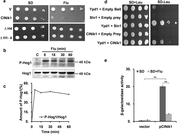
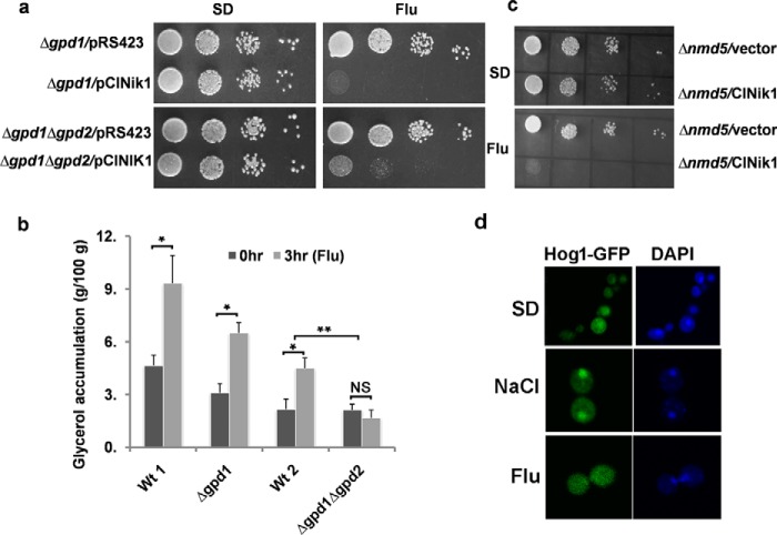
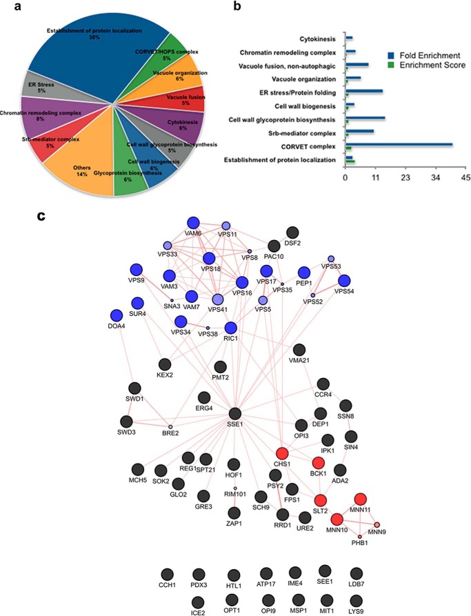
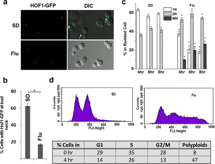
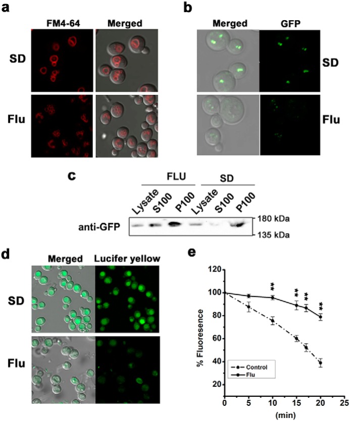
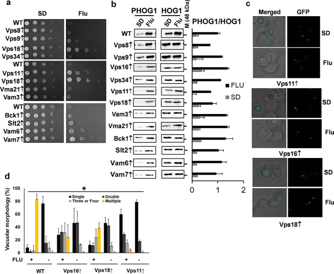
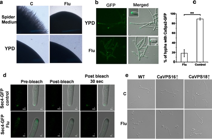
The hybrid histidine kinase 3 (HHK3) is a highly conserved sensor kinase in fungi that regulates the downstream HOG/p38 mitogen-activated protein kinase (MAPK). In addition to its role in osmoadaptation, HHK3 is involved in hyphal morphogenesis, conidiation, virulence, and cellular adaptation to oxidative stress. However, the molecular mechanisms by which it controls these processes remain obscure. Moreover, HHK3 is a molecular target for antifungal agents such as fludioxonil, which thereby interferes with the HOG/p38 pathway, leading to the abnormal accumulation of glycerol and subsequent cell lysis. Here, we used a chemical genomics approach with the yeast Saccharomyces cerevisiae to better understand the fungicidal action of fludioxonil and the role of HHK3 in fungal growth and physiology. Our results indicated that the abnormal accumulation of glycerol is not the primary cause of fludioxonil toxicity. Fludioxonil appears to impair endosomal trafficking in the fungal cells. We found that the components of class C core vacuole/endosome tethering (CORVET) complex are essential for yeast viability in the presence of a subthreshold dose of fludioxonil and that their overexpression alleviates fludioxonil toxicity. We also noted that by impeding secretory vesicle trafficking, fludioxonil inhibits hyphal growth in the opportunistic fungal pathogen Candida albicans Our results suggest that HHK3 regulates fungal hyphal growth by affecting vesicle trafficking. Together, our results reveal an important role of CORVET complex in the fungicidal action of fludioxonil downstream of HHK3.

Keywords: Candida albicans; HOG/p38 pathway; MAPK signaling; antifungal agent; cell signaling; filamentous growth; fludioxonil; fungi; histidine kinase; hybrid histidine kinase 3; mitogen-activated protein kinase (MAPK); p38 MAPK; vesicle trafficking; yeast.


Conflict of interest statement

The authors declare that they have no conflicts of interest with the contents of this article

Figures

Figure 1.
Figure 1.
ClNIK1 confers fludioxonil sensitivity to S. cerevisiae. a, dilution spotting of BY4741 harboring pRS423 (V), pClNIK1, or its mutants. 5 μl of a 10-fold serial dilution of exponential-phase cultures grown in SD medium were spotted on an SD agar plate containing 5 μg/ml fludioxonil (Flu). Plates were incubated at 28 °C for 2 days. b, immunoblot showing fludioxonil-induced Hog1p phosphorylation in S. cerevisiae strain BY4741 expressing ClNik1p. Cells were grown in SD minimal medium without histidine at 28 °C to logarithmic phase and treated with fludioxonil (5 μg/ml) for different times. Total cell extract from these samples was immunoblotted using anti-phospho-p38 antibody for phospho-Hog1p (P-Hog1) and anti-Hog1 antibody for total Hog1p (Hog1). C, control. c, graph showing the time course of Hog1p phosphorylation. Values are expressed as a percentage of Hog1p as obtained by densitometric analysis of Western blotting data. d, two-hybrid interactions of Ypd1p with Sln1p and ClNik1p. S. cerevisiae strain EGY48 transformed with different bait/prey constructs was grown in minimal SD medium, and a serial dilution of the culture was spotted onto a minimal SD and galactose-raffinose plate. e, in vivo histidine kinase activity of ClNik1p in the presence of fludioxonil. β-Gal activity in the yeast strain YSH2472 harboring pClNIK1 was determined after growth in minimal liquid medium in the absence (dark gray) or presence of 5 μg/ml fludioxonil (light gray). Experiments were repeated with two independent pools of six transformants each. β-Gal activity is expressed as nmol of o-nitrophenyl β-galactoside utilized/min by 1 ml of culture, which was normalized to an A600 of 1.0. The results are presented as mean ± S.D. (n = 9). **, p < 0.001. Error bars represent S.D.
Figure 2.
Figure 2.
Role of intracellular glycerol accumulation in the antifungal activity of fludioxonil. a, dilution spotting of S. cerevisiae strains Y13718 (Δgpd1) and 512 (Δgpd1Δgpd2) harboring vector pRS423 or pClNIK1 on an SD agar plate with or without 25 μg/ml fludioxonil (Flu). b, intracellular glycerol content of S. cerevisiae strains BY4742 (Wt 1), Y13718, 507 (Wt 2), and 512 harboring pClNIK1. Cultures at mid-exponential phase were exposed to 25 μg/ml fludioxonil for 3 h, and the amount of glycerol in total cell extract was measured using a UV glycerol assay kit (R-Biopharm) and normalized to dry weight of the cell. Results of two independent experiments performed in duplicates are presented as mean ± S.D. (n = 4). NS, not significant; *, p < 0.01; and **, p < 0.001. Error bars represent S.D. c, dilution spotting of S. cerevisiae strains B0119B (Δnmd5) harboring p426TEF (vector) or p426TEF-ClNIK1 (ClNik1) on an SD agar plate with or without 25 μg/ml fludioxonil. d, cellular localization of Hog1-GFP. Log-phase cultures of BY4741 harboring plasmids pClNIK1 and pHog1-GFP were treated with 0.4 m NaCl or 5 μg/ml fludioxonil for 15 min. After fixing cells with paraformaldehyde and staining with DAPI, GFP fluorescence in the untreated (SD) and treated samples was observed under a fluorescence confocal microscope.
Figure 3.
Figure 3.
GO enrichment of deletion strains hypersensitive to fludioxonil. a, pie chart describing the number and distribution of enriched GO terms obtained from DAVID analysis of fludioxonil deletion-sensitive profile. b, enrichment score and -fold enrichment of the identified GO terms. c, interaction network generated by GeneMANIA. Edges indicate physical interactions between gene products; edge weights reflect confidence in support for interactions. Blue node color corresponds to a set of functional gene classes involved in endosomal transport and vesicle fusion. Genes involved in cell wall biosynthesis are shown in red. Light-colored nodes were not part of the input set.
Figure 4.
Figure 4.
Effect of fludioxonil on the cell cycle. a, localization of Hof1-GFP in BY4741/pClNIK1. Cells at logarithmic phase were treated with fludioxonil (Flu) (25 μg/ml) for 3 h and observed under a microscope after fixing with paraformaldehyde. Localization of Hof1-GFP at the bud neck in the control untreated cells is shown by an arrow. DIC, differential interference contrast. b, graph showing the percentage of budded cells having Hof1-GFP localized at the bud neck. More than 100 cells were counted for each sample, and the data (mean ± S.D.) presented are from three independent experiments. *, p < 0.05. Error bars represent S.D. c, an early log-phase culture of BY4741/pClNIK1 synchronized by α-factor was released into fresh SD medium with or without 25 μg/ml fludioxonil. Cells were harvested at different times, stained with DAPI to monitor nuclear division, and observed under a microscope as described under “Experimental procedures.” The number of nuclei was counted in more than 100 budded cells in each sample (1N, 2N, and MN represent one, two, and three nuclei per cell). Results of three independent experiments are presented as mean ± S.D. *, p < 0.05. Error bars represent S.D. d, DNA content of S. cerevisiae strain BY4742/pClNIK1 with or without fludioxonil treatment was determined by flow cytometry as described under “Experimental procedures.” The x axis represents relative DNA content. The left-most peak represents the G1 population, and the right-most peak represents the G2 population. Representative data from three independent experiments are shown.
Figure 5.
Figure 5.
Effect of fludioxonil on vacuolar morphology and vesicle trafficking. a, S. cerevisiae strain BY4741/pClNIK1 was treated with 5 μg/ml fludioxonil (Flu) and stained with membrane dye FM4−64 as described under “Experimental procedures.” Micrographs were taken using a Nikon A1R confocal microscope. b, S. cerevisiae strain BY4741 with chromosomally tagged VPS8-GFP and harboring plasmid pClNIK1 was grown to log phase and treated with fludioxonil (5 μg/ml) for 2 h. Cells were washed with PBS and observed under a Nikon A1R confocal microscope. c, subcellular localization of GFP-tagged Vps8 was monitored in cells expressing ClNik1 after treating with fludioxonil (5 μg/ml) for 4 h. Total cell extract (Lysate) were fractionated to obtain a pellet (P100) and supernatant (S100) after the final centrifugation at 100,000 × g. Western blots were decorated against the GFP tag to identify Vps8. d, effect of fludioxonil on the accumulation of LY. S. cerevisiae strain BY4741/pClNIK1 with or without fludioxonil treatment was incubated at 30 °C with YPD containing LY (4 mg/ml) for 45 min before being photographed using a Nikon A1R confocal microscope. e, real-time FM4-64 recycling assay with S. cerevisiae strain BY4741/pClNIK1 treated with or without fludioxonil. FM4-64 fluorescence in the cell suspension was measured for 20 min continuously in a spectrofluorometer. Results are presented as mean ± S.D. (n = 6). **, p < 0.001. Error bars represent S.D.
Figure 6.
Figure 6.
Overexpression of the component of CORVET complex suppresses antifungal activity of fludioxonil. a, dilution spotting of S. cerevisiae strain BY4741 harboring different genes in multicopy plasmid pRS426 along with pClNIK1 on SD or an SD agar plate with fludioxonil (Flu) (25 μg/ml). b, immunoblot showing fludioxonil-induced Hog1p phosphorylation in S. cerevisiae strain BY4741 harboring different genes in multicopy plasmid pRS426 along with pClNIK1. The right panel shows the quantification of the phosphorylated Hog1p (PHOG1) in the immunoblot. The values were determined by densitometry and expressed as ratio of signals, PHOG1/HOG1, of the respective lanes. Graphs are plotted as mean ± S.D. calculated from three independent blots. Error bars represent S.D. c, localization of VPS8-GFP in S. cerevisiae strain BY4741/pClNIK1 overexpressing Vps11, Vps16, or Vps18. Cells at logarithmic phase were treated with fludioxonil (5 μg/ml) for 2 h. Cells were washed with PBS and observed under a Nikon A1R confocal microscope. A representative figure from three independent experiments is shown. d, vacuole fragmentation in S. cerevisiae strain BY4741/pClNIK1 overexpressing Vps11, Vps16, or Vps18. Cells at logarithmic phase untreated or treated with 5 μg/ml fludioxonil were visualized after staining with the dye FM4-64. Vacuoles with different morphology were counted and expressed as a percentage (mean ± S.D.) from three independent experiments (two-way analysis of variance; *, p < 0.05). Error bars represent S.D.
Figure 7.
Figure 7.
Fludioxonil inhibit hyphal growth in C. albicans. a, overnight cultures of C. albicans strain SC5314 were spotted onto YPD and a Spider agar plate with or without fludioxonil (Flu) (25 μg/ml) and incubated at 37 °C for 5 days. Images of colony edges were obtained using a stereomicroscope. C, control. b, fludioxonil affects the localization of CaSpa2p-GFP to the tips of filaments. Strain WYZ9 (Catup1Δ) expressing CaSpa2p-GFP was grown in YPD medium with or without 50 μg/ml fludioxonil at 37 °C for 6 h. Cells were washed with 1× PBS before visualizing under a microscope. c, percentage of hypha with CaSpa2p-GFP at the tip. The results are presented as mean ± S.D. (n = 3). **, p < 0.01. Error bars represent S.D. d, fluorescence recovery of CaSec4-GFP. C. albicans strain expressing CaSec4-GFP was grown in Spider medium at 37 °C for 45 min. After addition of fludioxonil (50 μg/ml), the culture was incubated further for 45 min. Cells were then embedded in 0.4% agar on glass slides for FRAP. A minimum of 10 hyphal tips of each strain were bleached, and images were recorded before bleaching (prebleach) for 30 s, 30 s after bleaching (postbleach), and 60s after bleaching. Representative images of individual hyphal tips at prebleach, postbleach, and 30 s postbleach are shown. Bars, 1 μm. e, hyphal morphology of C. albicans strains overexpressing CaVPS16 and CaVPS18. ORFs encoding CaVPS16 and CaVPS18 were cloned in plasmid pVT50 under the ACT1 promoter, and the linearized plasmids were chromosomally integrated into C. albicans strain SC5314 at the RP10 (ribosomal protein 10) gene locus. Cells were grown in Spider medium for 3 h at 37 °C with and without fludioxonil (25 μg/ml) and observed under a microscope. C. albicans strain SC5314 was used as a control (WT).

Similar articles

Cited by

References

    1. Brown G. D., Denning D. W., Gow N. A., Levitz S. M., Netea M. G., and White T. C. (2012) Hidden killers: human fungal infections. Sci. Transl. Med. 4, 165rv13 10.1126/scitranslmed.3004404 - DOI - PubMed
    1. Denning D. W., and Bromley M. J. (2015) How to bolster the antifungal pipeline. Science 347, 1414–1416 10.1126/science.aaa6097 - DOI - PubMed
    1. Scorzoni L., de Paula E. Silva A. C., Marcos C. M., Assato P. A., de Melo W. C., de Oliveira H. C., Costa-Orlandi C. B., Mendes-Giannini M. J., and Fusco-Almeida A. M. (2017) Antifungal therapy: new advances in the understanding and treatment of mycosis. Front. Microbiol. 8, 36 10.3389/fmicb.2017.00036 - DOI - PMC - PubMed
    1. Myung K., and Klittich C. J. (2015) Can agricultural fungicides accelerate the discovery of human antifungal drugs? Drug Discov. Today 20, 7–10 10.1016/j.drudis.2014.08.010 - DOI - PubMed
    1. Corran A., Knauf-Beiter G., and Zeun R. (2008) Fungicides acting on signal transduction, in Modern Crop Protection Compounds (Krämer W., and Schirmer U., eds) pp. 561–580, Wiley-VCH Verlag GmbH, Weinheim, Germany

Publication types

MeSH terms

LinkOut - more resources