. 2018 Dec 12;145(24):dev168922.
doi: 10.1242/dev.168922.

The RhoGEF protein Plekhg5 regulates apical constriction of bottle cells during gastrulation

Affiliations

The RhoGEF protein Plekhg5 regulates apical constriction of bottle cells during gastrulation

Ivan K Popov et al. Development. .

Abstract

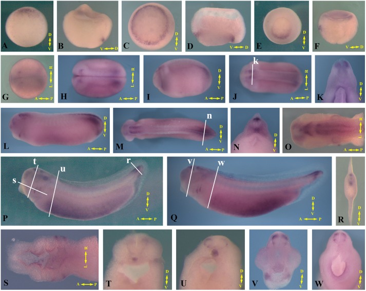
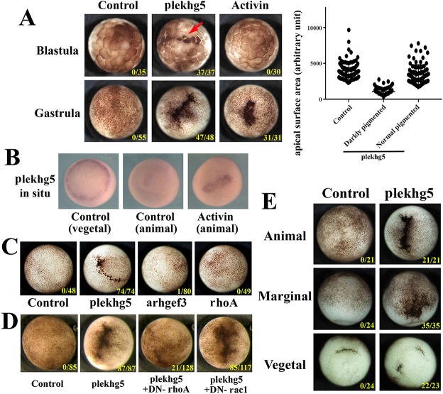
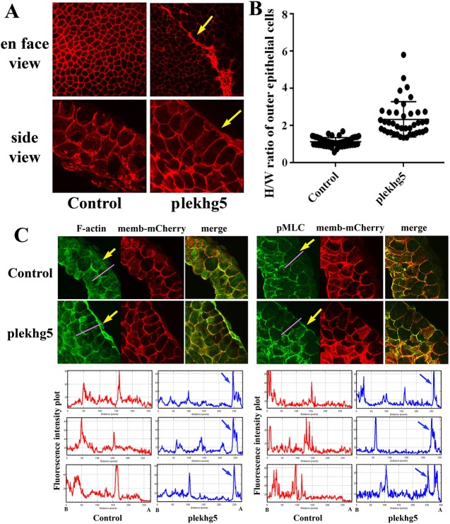
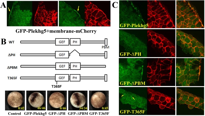
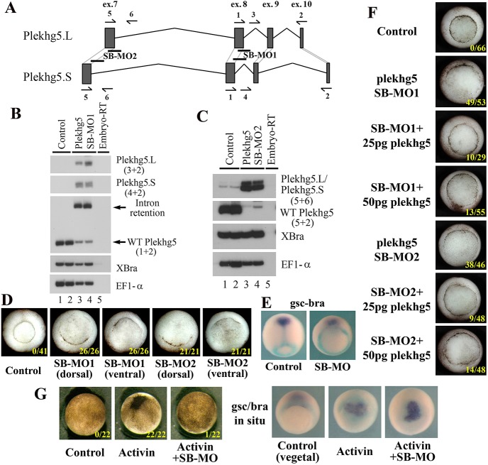
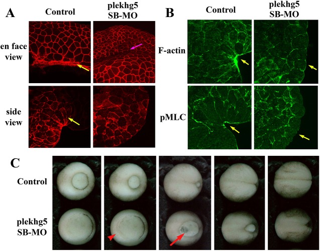
Apical constriction regulates epithelial morphogenesis during embryonic development, but how this process is controlled is not understood completely. Here, we identify a Rho guanine nucleotide exchange factor (GEF) gene plekhg5 as an essential regulator of apical constriction of bottle cells during Xenopus gastrulation. plekhg5 is expressed in the blastopore lip and its expression is sufficient to induce ectopic bottle cells in epithelia of different germ layers in a Rho-dependent manner. This activity is not shared by arhgef3, which encodes another organizer-specific RhoGEF. Plekhg5 protein is localized in the apical cell cortex via its pleckstrin homology domain, and the GEF activity enhances its apical recruitment. Plekhg5 induces apical actomyosin accumulation and cell elongation. Knockdown of plekhg5 inhibits activin-induced bottle cell formation and endogenous blastopore lip formation in gastrulating frog embryos. Apical accumulation of actomyosin, apical constriction and bottle cell formation fail to occur in these embryos. Taken together, our data indicate that transcriptional regulation of plekhg5 expression at the blastopore lip determines bottle cell morphology via local polarized activation of Rho by Plekhg5, which stimulates apical actomyosin activity to induce apical constriction.

Keywords: Actin; Apical constriction; Bottle cell; Gastrulation; Myosin; RhoGEF; Xenopus.


Conflict of interest statement

Competing interestsThe authors declare no competing or financial interests.

Figures

Fig. 1.
Fig. 1.
Dynamic expression of plekhg5 in early Xenopus embryos. (A-F) plekhg5 is expressed in the blastopore lip during gastrulation. Vegetal view (A,C,E) and side view of bisected embryos (B,D,F) are shown. (G-O) During neurula (G,H) and tailbud (I-O) stages, plekhg5 is expressed in the tail, hindbrain, otic and olfactory placodes, and pharyngeal pouch. Sections of the embryos reveal plekhg5 transcripts in the dorsal neural tube and the notochord. (P,Q) Expression of plekhg5 at the tadpole stages is detected in the hindbrain, otic vesicles, tail, pharyngeal pouch and the ventral-lateral mesoderm. (R-W) Sections of the tadpole embryos show expression of plekhg5 in the notochord, transiently in the trunk but persisting in the tail, the migrating neural crest cells along the ventral route and in the dorsal root ganglia, the dorsal neural tube in the tail, the tips of the outgrowing pharyngeal pouches, and the lining of the foregut. White lines in J,M,P,Q show the position of the sections in the panels indicted by the letters. The embryonic axes are labeled in each panel: D-V, dorsal-ventral; A-P, anterior-posterior; L-R, left-right.
Fig. 2.
Fig. 2.
plekhg5 induces ectopic blastopore lip-like morphology in early Xenopus embryos in a Rho-dependent manner. (A) plekhg5 expression induces apical cell constriction in ectodermal cells at early blastula stages, whereas activin induces ectopic blastopore lip at the gastrula stages. The apical surface areas of cells at the blastula stages in control and the plekhg5-expressing embryos are measured and compared. The scatter plot shows a typical experiment. plekhg5 significantly reduces apical cell surfaces to about one-third that seen in control cells, with the average surface areas of 3978, 1050 and 3409 (arbitrary units) for control, apically constricted, and normal pigmented cells, respectively. Student's t-test gives a P-value of 3.5E-31 in this experiment. Red arrow indicates apically constricted cells at the blastula stages. (B) Activin induces expression of plekhg5 in the ectoderm when it induces an ectopic blastopore lip. (C) Unlike plekhg5, neither arhgef3 nor general expression of rhoA induces ectopic blastopore lip morphology in the ectoderm. (D) Dominant-negative rhoA, but not rac1, blocks ectopic blastopore lip induction by plekhg5. (E) plekhg5 induces ectopic blastopore lip morphology when injected either in the animal, the marginal zone or the vegetal regions. The doses of RNAs used are 100 pg of plekhg5, 5 pg of activin, 200 pg arhgef3, 0.5-1 ng of rhoA, DN-rhoA and DN-rac1. Numbers in each image indicate embryos exhibiting the ectopic blastopore lip-like morphology over the total number of embryos. All the experiments are repeated at least three times.
Fig. 3.
Fig. 3.
plekhg5 induces cell elongation and apical actomyosin accumulation in outer epithelial cells. (A) En face view of early gastrula embryos shows reduced cell surfaces in plekhg5-expressing cells (arrows) compared with those in control embryos. Side view of the bisected embryos shows elongation of superficial epithelial cells from plekhg5-injected embryos. (B) H/W ratio analysis shows that plekhg5-expressing outer epithelial cells have a significant increase in H/W ratio from 1.2 in control cells to 2.2 in plekhg5-expressing cells. Student's t-test gives P=8.3E-21. (C) plekhg5 stimulates apical accumulation of both F-actin and pMLC. The membrane-mCherry signal is used to label the injected cells. Arrows indicate the apical F-actin and pMLC signals. Fluorescence intensity is measured using ImageJ along the axis indicated by the pink line across the animal regions. The plots for three different biological samples are shown with the apical (A) and basal (B) direction labeled at the bottom. Blue arrows point to the apical enhancement of F-actin and pMLC signals.
Fig. 4.
Fig. 4.
Plekhg5 is apically localized in the superficial epithelial cells. (A) GFP-tagged Plekhg5 protein is detected at the apical cell cortex in the superficial epithelial cells (arrows), but is diffuse in deeper ectodermal cells. (B) Plekhg5 contains a PH domain and a PBM in addition to the GEF domain. Analyses of the deletion mutants that lack one of these domains reveal that removal of the PH domain, but not the PBM motif, abolishes the ability of the protein to induce ectopic blastopore lip. In addition, a point mutation that alters the conserved threonine 365 residue in the GEF domain into phenylalanine also results in non-functional Plekhg5. Numbers in each image indicate embryos exhibiting the ectopic blastopore lip-like morphology over the total number of embryos. (C) Deletion of the PH domain, but not the PBM, results in loss of apical accumulation of the proteins. The T365F GEF mutant protein can be recruited to the cell junctions in epithelial cells (arrows), but is not enriched at the apical cell cortex.
Fig. 5.
Fig. 5.
plekhg5 is required for endogenous blastopore lip formation. (A) Schematic of the genomic regions of the L and the S alloalleles of plekhg5 that are targeted by the SB MOs. The positions of the primers used for RT-PCR analysis of splicing efficiency are shown. (B,C) Both SB-MO1 and SB-MO2 efficiently block splicing of both L and S alloalleles, as indicated by the presence of intron-retention products in plekhg5 morphant embryos. The primer pairs used in the PCR reactions are indicated in parentheses. (D) plekhg5 SB MOs prevent formation of the blastopore lip at the sites of its injection. (E) plekhg5 SB MOs do not alter mesodermal cell fates, though the movements of the prechordal tissue (gsc-expressing, purple) and the trunk mesoderm (bra-expressing, cyan) are affected. (F) The blastopore lip defects induced by the SB MOs (25 ng) can be rescued with low doses of co-expressed plekhg5 RNA (25-50 pg). (G) plekhg5 SB MOs (25 ng) block ectopic blastopore lip induction by activin (5 pg) without affecting activin-dependent mesodermal induction. Numbers in each image indicate embryos exhibiting the ectopic blastopore lip defects or ectopic blastopore lips over the total number of embryos.
Fig. 6.
Fig. 6.
plekhg5 regulates apical actomyosin cytoskeleton in bottle cells and gastrulation movements. (A) En face and side views of control bottle cells show reduced cell surfaces and wedge-shaped morphology in gastrula embryos, respectively (yellow arrows). However, in plekhg5 morphant embryos, cells do not show great shrinkage of surface areas and only cuboidal epithelial cell shapes are seen from the side view. Despite this, internalization of surface cells appears to happen at imprecise positions in the morphant embryos, as shown by formation of a surface groove (pink arrow). (B) Both F-actin and pMLC are enriched in the apical cell cortex of the bottle cells in bisected control embryos, but no such enrichment is observed in plekhg5 morphant embryos. Yellow arrows indicate apical signals. (C) Gastrulation movements proceed in the absence of the bottle cells, as seen by accumulation of cells in the marginal region from epiboly (red arrowhead) and thinning of the vegetal mass due to rotational movements of the large endodermal cells upward and laterally (red arrow). The blastopore eventually closes in most morphant embryos, but is delayed, when control siblings reach the neurula stages. Selected still frames from a time-lapse video of gastrulating control and plekhg5 morphant embryos are shown.

References

    1. Aghazadeh B., Zhu K., Kubiseski T. J., Liu G. A., Pawson T., Zheng Y. and Rosen M. K. (1998). Structure and mutagenesis of the Dbl homology domain. Nat. Struct. Biol. 5, 1098-1107. 10.1038/4209 - DOI - PubMed
    1. Anderson D. C., Gill J. S., Cinalli R. M. and Nance J. (2008). Polarization of the C elegans embryo by RhoGAP-mediated exclusion of PAR-6 from cell contacts. Science 320, 1771-1774. 10.1126/science.1156063 - DOI - PMC - PubMed
    1. Barmich M. J., Rogers S. and Häcker U. (2005). DRhoGEF2 regulates actin organization and contractility in the Drosophila blastoderm embryo. J. Cell Biol. 168, 575-585. 10.1083/jcb.200407124 - DOI - PMC - PubMed
    1. Barrett K., Leptin M. and Settleman J. (1997). The Rho GTPase and a putative RhoGEF mediate a signaling pathway for the cell shape changes in Drosophila gastrulation. Cell 91, 905-915. 10.1016/S0092-8674(00)80482-1 - DOI - PubMed
    1. Baumeister M. A., Rossman K. L., Sondek J. and Lemmon M. A. (2006). The Dbs PH domain contributes independently to membrane targeting and regulation of guanine nucleotide-exchange activity. Biochem. J. 400, 563-572. 10.1042/BJ20061020 - DOI - PMC - PubMed

Publication types

MeSH terms