. 2018 Dec 10;145(24):dev158352.
doi: 10.1242/dev.158352.

Spatiotemporal control of axillary meristem formation by interacting transcriptional regulators

Affiliations

Spatiotemporal control of axillary meristem formation by interacting transcriptional regulators

Cui Zhang et al. Development. .

Abstract

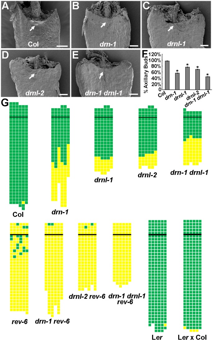
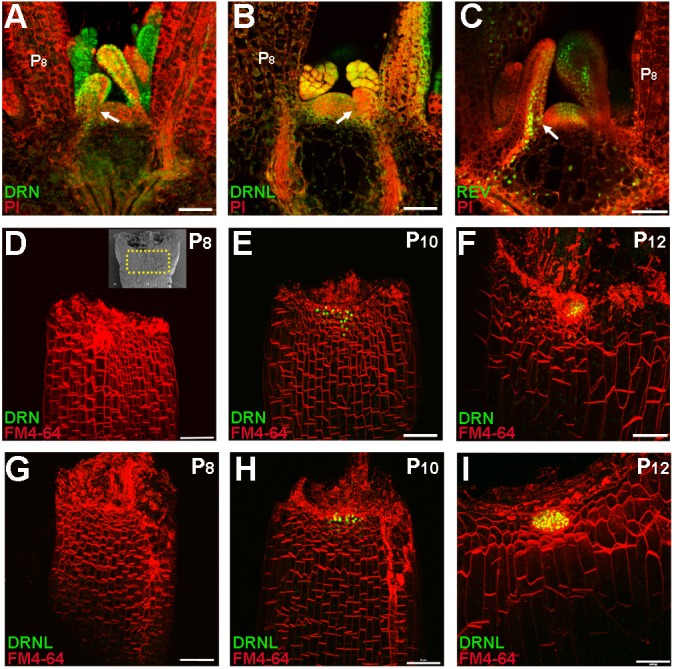
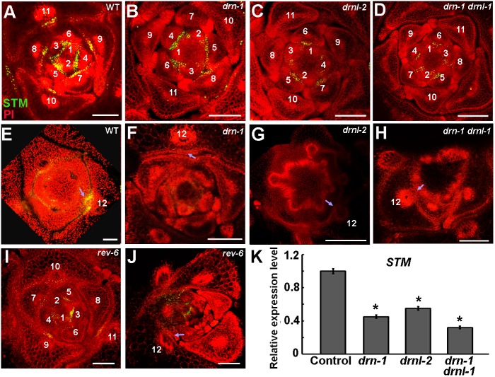
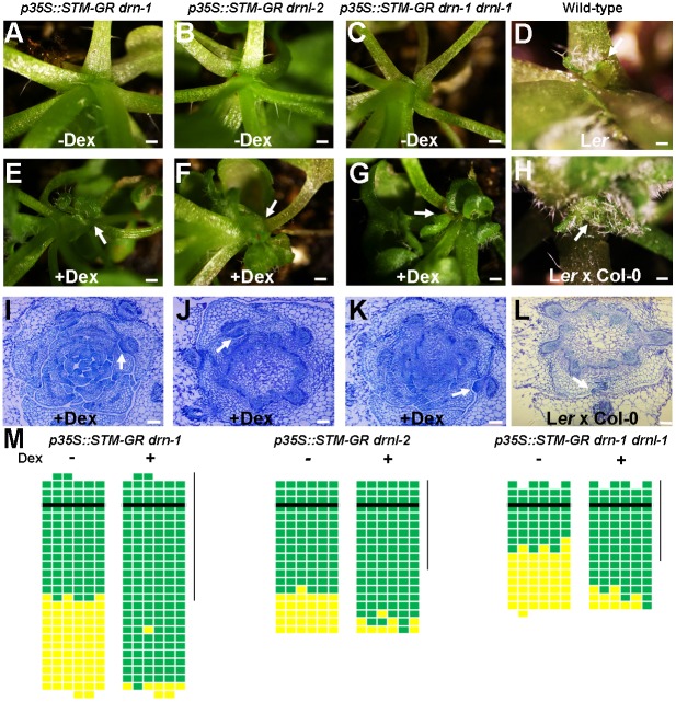
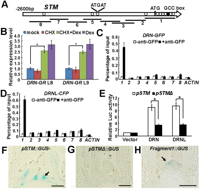
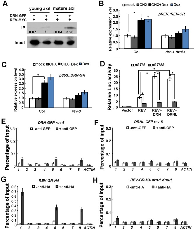
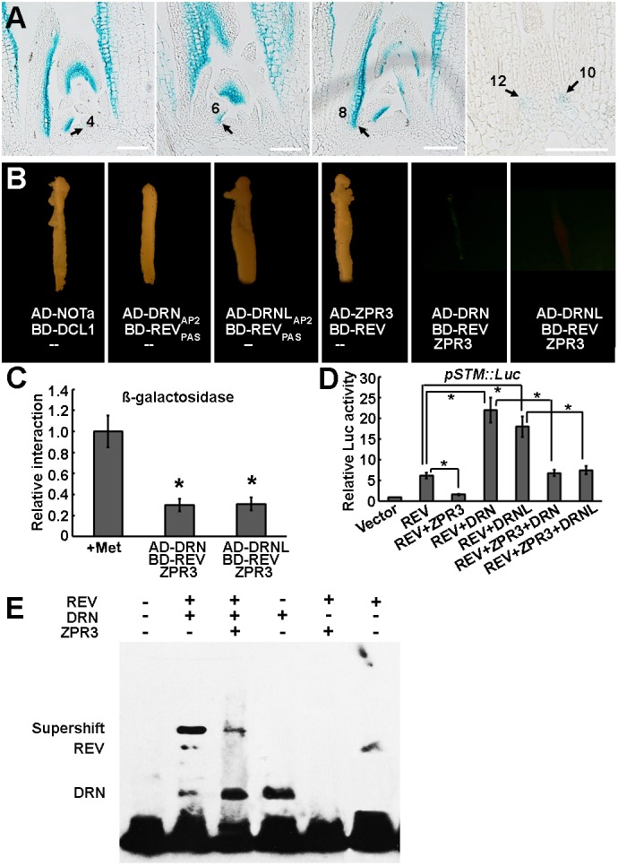
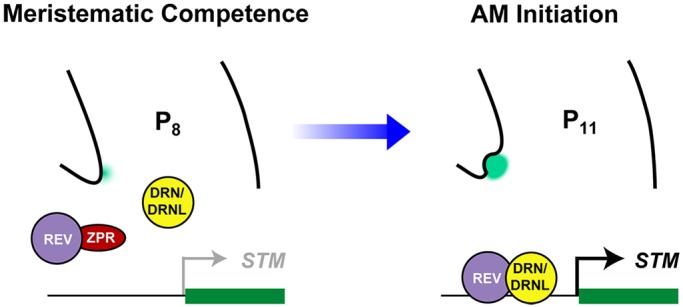
Branching is a common feature of plant development. In seed plants, axillary meristems (AMs) initiate in leaf axils to enable lateral shoot branching. AM initiation requires a high level of expression of the meristem marker SHOOT MERISTEMLESS (STM) in the leaf axil. Here, we show that modules of interacting transcriptional regulators control STM expression and AM initiation. Two redundant AP2-type transcription factors, DORNRÖSCHEN (DRN) and DORNRÖSCHEN-LIKE (DRNL), control AM initiation by regulating STM expression. DRN and DRNL directly upregulate STM expression in leaf axil meristematic cells, as does another transcription factor, REVOLUTA (REV). The activation of STM expression by DRN/DRNL depends on REV, and vice versa. DRN/DRNL and REV have overlapping expression patterns and protein interactions in the leaf axil, which are required for the upregulation of STM expression. Furthermore, LITTLE ZIPPER3, another REV-interacting protein, is expressed in the leaf axil and interferes with the DRN/DRNL-REV interaction to negatively modulate STM expression. Our results support a model in which interacting transcriptional regulators fine-tune the expression of STM to precisely regulate AM initiation. Thus, shoot branching recruits the same conserved protein complexes used in embryogenesis and leaf polarity patterning.

Keywords: Arabidopsis; Axillary meristem; Branching; Stem cell; Transcription.


Conflict of interest statement

Competing interestsThe authors declare no competing or financial interests.

Figures

Fig. 1.
Fig. 1.
DRN and DRNL are required for AM initiation. (A-E) Scanning electron micrographs of P15 rosette leaf axils in Col-0 wild type with a developing AM (dense cell mass, arrow) (A), and in drn-1 (B), drnl-1 (C), drnl-2 (D) and drn-1 drnl-1 (E) mutants with bare axils (arrows). (F) Axillary bud formation in drn-1, drnl-1, drnl-2 and drn-1 drnl-1 mutants during vegetative development in comparison with Col-0 wild-type plants. The percentage values indicate the mean proportion of axillary buds formed over the total number of leaves along the shoot axis (plants analyzed >20). Error bars indicate the s.d. *P<0.01 between wild type and each mutant. (G) Schematic representation of axillary bud formation in leaf axils of Col-0 wild-type plants; drn-1, drnl-1, drnl-2, drn-1 drnl-1, rev-6, drn-1 rev-6, drnl-2 rev-6 and drn-1 drnl-1 rev-6 mutant plants; Ler, and mixed Ler and Col-0 ecotypes. The thick black horizontal line represents the border between the youngest rosette leaf and the oldest cauline leaf. Each column represents a single plant, and each square within a column represents an individual leaf axil. The bottom row represents the oldest rosette leaf axils, with progressively younger leaves above. Green indicates the presence of an axillary bud; yellow indicates the absence of an axillary bud in any particular leaf axil. Scale bars: 100 μm.
Fig. 2.
Fig. 2.
DRN and DRNL are expressed in leaf primordia and accumulate in the leaf axil prior to AM initiation. (A-C) Expression of pDRN::DRN-GFP (A), pDRNL::DRNL-CFP (B) and pREV::REV-Venus (C) in the vegetative shoot apex and leaf primordia. Longitudinal sections of 14-day-old plant shoot apices were stained with propidium iodide (PI, red); fluorescent signals are shown in green. Arrows indicate leaf axils. Fluorescent signals are present in the leaf axils. (D-I) Reconstructed view of the epidermal layer of P8 (D,G), P10 (E,H) and P12 (F,I) leaf axils with pDRN::DRN-GFP (D-F) or pDRNL::DRNL-CFP (G-I) expression in green and FM4-64 staining in red showing the location of AM progenitor cells. The inset in D shows a scanning election micrograph of a rosette leaf axil at a similar stage; the region within the yellow dotted box roughly corresponds to the imaged regions shown in D-I. All leaves were removed from 17-day-old plants. Note the enrichment of DRN-GFP and DRNL-CFP signals in P10 and P12 leaf axils. Scale bars: 50 μm.
Fig. 3.
Fig. 3.
Attenuated STM expression in drn and drnl mutants. (A-D) Patterns of pSTM::STM-Venus (green) in transverse sections through the vegetative shoot apex of 28-day-old wild-type Col-0 (A), drn-1 (B), drnl-2 (C) and drn-1 drnl-1 (D) plants. Plants are stained with propidium iodide (PI, red). The STM expression level is decreased in the mutants compared with wild type. (E-H) Patterns of pSTM::STM-Venus expression in P12 leaf axils in wild-type Col-0 (E), drn-1 (F), drnl-2 (G) and drn-1 drnl-1 (H). STM expression levels are lower in mature leaves in the mutants than in wild type. (I,J) STM-Venus expression levels in rev-6 in young (I) and mature (J) leaves. Arrows indicate leaf axils; numbers in A-J indicate leaf stages. (K) RT-qPCR analysis indicates that STM expression is significantly reduced in drn-1, drnl-2 and drn-1 drnl-1 mutant plants. Vegetative shoots with the leaves removed were analyzed. Error bars indicate s.d. *P<0.01 (Student's t-test). Scale bars: 100 μm.
Fig. 4.
Fig. 4.
Overexpression of STM rescues axillary bud deficiency in drn and drnl mutants. (A-C) Higher magnification of rosette leaf axils in mock-treated p35S::STM-GR drn-1 (A), p35S::STM-GR drnl-2 (B) and p35S::STM-GR drn-1 drnl-1 (C) plants showing the absence of an axillary bud. (E-G) Higher magnification of rosette leaf axils in Dex-treated p35S::STM-GR drn-1 (E), p35S::STM-GR drnl-2 (F) and p35S::STM-GR drn-1 drnl-1 (G) plants showing the presence of axillary buds (arrows). (D,H) Higher magnification of rosette leaf axils in mock-treated Ler and mixed Ler×Col-0 ecotypes showing the presence of axillary buds (arrows). (I-L) Transverse sections through vegetative shoot apices of 28-day-old Dex-treated p35S::STM-GR drn-1 (I), p35S::STM-GR drnl-2 (J), p35S::STM-GR drn-1 drnl-1 (K) and mixed Ler×Col-0 ecotypes (L) stained with Toluidine Blue O, showing the presence of axillary buds (arrows) in rosette leaf axils. (M) Schematic representation of axillary buds in leaf axils with or without Dex induction. Green indicates the presence of an axillary bud; yellow indicates the absence of an axillary bud. Plants were grown under short-day conditions for 15 days without treatment; leaf axil regions were treated with 10 µM Dex every second day for another 15 days and then transferred to long-day conditions without treatment until axillary buds were counted. The vertical line indicates leaves initiated during Dex treatment. Horizontal lines indicate the border between the youngest rosette leaf and the oldest cauline leaf. Scale bars: 2 cm in A-H; 100 μm in I-L.
Fig. 5.
Fig. 5.
DRN and DRNL regulate STM expression via binding to a conserved promoter motif. (A) Schematic representation of the STM genomic region. The black circle and white circles indicate the GCCGCC motif and the ATGAT motif, respectively; ATG denotes the translation start site. Eight PCR fragments were designed for ChIP analysis. (B) RT-qPCR analysis of STM expression using p35S::DRN-GR vegetative shoots (with the leaves removed) before and after simultaneous Dex and CHX treatment for 2 h. The vertical axis indicates the relative mRNA amount compared with the amount in the mock treatment. Error bars indicate the s.d. Two independent transgenic lines were used. *P<0.01 (Student's t-test). (C,D) ChIP-qPCR analysis indicates binding of DRN-GFP (C) and DRNL-CFP (D) to fragment 1. Error bars indicate the s.d. (E) Relative Luc reporter gene expression in transcriptional activity assays in Arabidopsis protoplasts. The 3.0 kb STM promoter region (pSTM) or the same region without fragment 1 (as indicated in A, pSTMΔ) was co-transformed with p35S::DRN or p35S::DRNL, and p35S::GUS was the internal control. Data are mean±s.d. for three independent biological experiments, each performed in triplicate. *P<0.01 (Student's t-test). (F-H) Pattern of GUS expression driven by pSTM (F), pSTMΔ (G) and fragment 1 (H) in longitudinal sections through a vegetative shoot apex of 30-day-old plants. To compare signals, plants were stained in parallel for 6 h, and sections were placed on the same slides for detection. The GUS signal is barely detectable in leaf axils of pSTMΔ::GUS plants (F) but weakly detectable in fragment1::GUS (H). Arrows highlight leaf axils. See Fig. S7 for more examples. Scale bars: 100 μm in F-H.
Fig. 6.
Fig. 6.
DRN and DRNL enhance the activation of STM expression by REV. (A) Co-IP assay indicating that DRN and REV interact in vivo and the interaction is stronger in mature leaf (older than P10) axils than in young leaf (younger than P10) axils. The numbers below the blots indicate the relative ratios of the signal intensity between IP and input bands (IP/input). The ratios were normalized to the IP band in young leaf axils of crossed marker lines. An anti-GFP antibody was used for IP and an anti-MYC antibody as a probe. Input shows the amount of DRN-GFP protein used in the IP assay. (B) RT-qPCR analysis of STM expression in pREV::REV-GR and pREV::REV-GR drn-1 drnl-1 vegetative shoots (with the leaves removed) before and after simultaneous Dex and CHX treatment for 2 h. The vertical axis indicates the relative mRNA amount compared with the amount in the mock treated. Error bars indicate the s.d. STM activation is reduced in pREV::REV-GR drn-1 drnl-1. *P<0.01 (Student's t-test). (C) RT-qPCR analysis of STM expression in p35S::DRN-GR and p35S::DRN-GR rev-6 vegetative shoots (with the leaves removed) before and after simultaneous Dex and CHX treatment for 2 h. The vertical axis indicates the relative mRNA amount compared with the amount in the mock treatment. Error bars indicate s.d. STM activation is reduced in p35S::DRN-GR rev-6. *P<0.01 (Student's t-test). (D) Relative Luc reporter gene expression in transcriptional activity assays in Arabidopsis protoplasts. The pSTM::Luc or the pSTMΔ::Luc constructs were co-transformed with p35S::REV alone, p35S::REV and p35S::DRN, or p35S::REV and p35S::DRNL; p35S::GUS was the internal control. Data are mean±s.d. Error bars are derived from three independent biological experiments, each performed in triplicate. Note the enhanced activation of STM expression by DRN and DRNL. *P<0.01 (Student's t-test). (E,F) ChIP-qPCR analysis demonstrates the reduced binding of DRN-GFP (E) and DRNL-CFP (F) to the STM genomic region (as in Fig. 5A) in rev-6 plants. Compare binding with that in Fig. 5C,D for Col-0 wild-type plants. Error bars indicate the s.d. (G,H) ChIP-qPCR analysis demonstrates binding of REV-GR-HA to the STM genomic region in Col-0 wild-type plants (G); this is reduced in drn-1 drnl-1 plants (H). Error bars indicate s.d.
Fig. 7.
Fig. 7.
ZPR3 interferes with the DRN/DRNL-REV interaction and inhibits STM expression. (A) Patterns of ZPR3-promoter-driven GUS expression in serial longitudinal 8 μm sections through the vegetative shoot apex of a 30-day-old wild-type-like plant. Arrows indicate the leaf axils. The GUS signals are weaker in the leaf axils of P10 and older leaves. See Fig. S8D-G for additional transverse sections. Scale bars: 100 μm. (B) Y2H and Y3H assay showing the disruption of the DRN/DRNL-REV interaction by ZPR3. Yeast growth on SD-Leu-Trp-His-Ade plates showing that DRN, DRNL and ZPR3 interact with REV, respectively. The interaction of DRN or DRNL with REV was weakened after the induction of ZPR3 activity. AD-NOTa and BD-DCL1 were used as positive controls. (C) Relative β-galactosidase activity of the UAS-driven β-galactosidase reporter measured before and after ZPR3 induction in Y3H. Constructs and additional results are shown in Fig. S9. The data are mean values of three replicates±s.d. *P<0.01 (Student's t-test). (D) Relative Luc reporter gene expression in transcriptional activity assays in Arabidopsis protoplasts. The pSTM::Luc construct was co-transformed with p35S::REV alone, p35S::REV+p35S::ZPR3, p35S::REV+p35S::DRN, p35S::REV+p35S::DRNL, p35S::REV+p35S::ZPR3+p35S::DRN or p35S::REV+p35S::ZPR3+p35S::DRNL; p35S::GUS was the internal control. Data are mean±s.d. Error bars are derived from three independent biological experiments, each performed in triplicate. Note the suppression of REV activation and DRN/DRNL–REV co-activation of STM expression by ZPR3. *P<0.01 (Student's t-test). (E) Supershift in EMSA, indicating that REV and DRN interact and bind to a biotin-labeled STM promoter fragment. The addition of ZPR3 decreased the intensity of the supershift band of DRN and REV; 2 µg DRN and ZPR3, and 1 µg REV protein were used for incubation.
Fig. 8.
Fig. 8.
A model summarizing the upregulation of STM expression prior to AM initiation through the licensing of active DRN/DRNL-REV complexes by ZPRs. In the early leaf axil, ZPR3 is expressed and interacts with REV to inhibit the formation of functional DRN/DRNL-REV complexes, resulting in a low level of STM expression and the absence of axillary bud formation. As the leaf matures, decreasing ZPR3 levels allow the formation of DRN/DRNL-REV complexes in more mature leaf axils to upregulate STM expression and to promote axillary bud formation.

References

    1. Aguilar-Martínez J. A., Uchida N., Townsley B., West D. A., Yanez A., Lynn N., Kimura S. and Sinha N. (2015). Transcriptional, posttranscriptional, and posttranslational regulation of SHOOT MERISTEMLESS gene expression in Arabidopsis determines gene function in the shoot apex. Plant Physiol. 167, 424-442. 10.1104/pp.114.248625 - DOI - PMC - PubMed
    1. Banno H., Ikeda Y., Niu Q. W. and Chua N. H. (2001). Overexpression of Arabidopsis ESR1 induces initiation of shoot regeneration. Plant Cell 13, 2609-2618. 10.1105/tpc.13.12.2609 - DOI - PMC - PubMed
    1. Banno H., Mase H. and Maekawa K. (2006). Analysis of functional domains and binding sequences of Arabidopsis transcription factor ESR1. Plant Biotechnol. 23, 303-308. 10.5511/plantbiotechnology.23.303 - DOI
    1. Brady S. M., Zhang L., Megraw M., Martinez N. J., Jiang E., Yi C. S., Liu W., Zeng A., Taylor-Teeples M., Kim D. et al. (2011). A stele-enriched gene regulatory network in the Arabidopsis root. Mol. Syst. Biol. 7, 459 10.1038/msb.2010.114 - DOI - PMC - PubMed
    1. Brand U., Grunewald M., Hobe M. and Simon R. (2002). Regulation of CLV3 expression by two homeobox genes in Arabidopsis. Plant Physiol. 129, 565-575. 10.1104/pp.001867 - DOI - PMC - PubMed

Publication types

MeSH terms