. 2018 Nov 16;9(1):4830.
doi: 10.1038/s41467-018-07378-5.

Dna2 processes behind the fork long ssDNA flaps generated by Pif1 and replication-dependent strand displacement

Affiliations

Dna2 processes behind the fork long ssDNA flaps generated by Pif1 and replication-dependent strand displacement

Silvia Emma Rossi et al. Nat Commun. .

Abstract

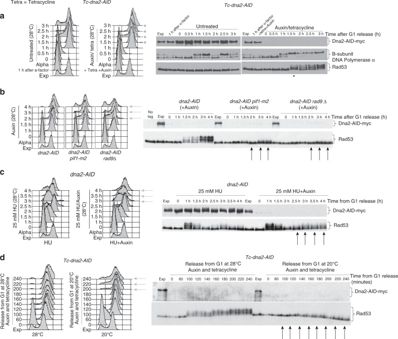
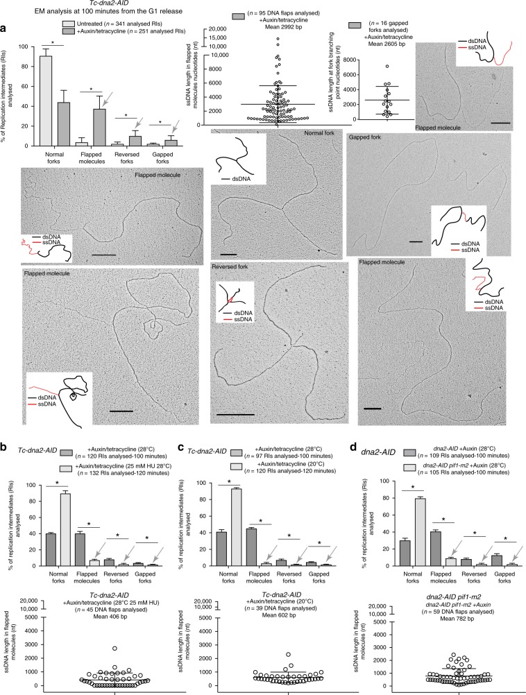
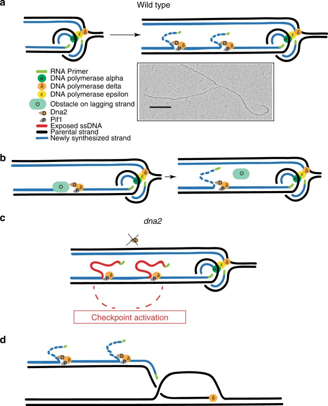
Dna2 is a DNA helicase-endonuclease mediating DSB resection and Okazaki fragment processing. Dna2 ablation is lethal and rescued by inactivation of Pif1, a helicase assisting Okazaki fragment maturation, Pol32, a DNA polymerase δ subunit, and Rad9, a DNA damage response (DDR) factor. Dna2 counteracts fork reversal and promotes fork restart. Here we show that Dna2 depletion generates lethal DNA structures activating the DDR. While PIF1 deletion rescues the lethality of Dna2 depletion, RAD9 ablation relieves the first cell cycle arrest causing genotoxicity after few cell divisions. Slow fork speed attenuates DDR in Dna2 deprived cells. Electron microscopy shows that Dna2-ablated cells accumulate long ssDNA flaps behind the forks through Pif1 and fork speed. We suggest that Dna2 offsets the strand displacement activity mediated by the lagging strand polymerase and Pif1, processing long ssDNA flaps to prevent DDR activation. We propose that this Dna2 function has been hijacked by Break Induced Replication in DSB processing.


Conflict of interest statement

The authors declare no competing interests.

Figures

Fig. 1
Fig. 1
Pif1 and fork speed induce RAD9-dependent DDR hyper-activation in Dna2-depleted CL-dna2 cells. ad Dna2 was depleted in G1 in the indicated strains and cells were released into unperturbed S phase (a, b), S phase in the presence of a low dose of HU (c) or at a low temperature (d). ac Non-depleted cells were kept in parallel as control. DNA content and the phosphorylation state of the indicated proteins were analysed, respectively,  by fluorescence-activated cell sorting (FACS) and western blotting at the indicated time points and in the specified genetic backgrounds. a Black asterisk and white circles indicate, respectively, Rad53 hyper-activation and first cell cycle arrest in Dna2-depleted cells. bd Black arrows and grey arrows indicate, respectively, lack of Rad53 activation and cells that initiate the second cell cycle
Fig. 2
Fig. 2
Fork speed and Pif1 induce cell death in Dna2-depleted cells. a In-plate cell survival assay. The indicated yeast strains were depleted (auxin) or non-depleted (untreated) for Dna2. Black and grey arrows indicate suppression of cell lethality induced by pif1-m2 mutation or RAD9 deletion. b In liquid growth curve of dna2-AID rad9Δ cells depleted for Dna2 in G1 (auxin) or non-depleted (untreated) and released into the cell cycle. The black arrow indicates growth arrest in dna2-AID rad9Δ cells depleted for Dna2. c, d In-plate cell survival assay. Black arrows indicate that low HU dose or low temperature suppress the cell lethality induced by depletion of Dna2 in dna2-AID rad9Δ cells. e Same experiment as in (b), but cells were released at 20 °C. In this condition, dna2-AID rad9Δ cells depleted for Dna2 do not arrest cell growth even after 15 h from the G1 release
Fig. 3
Fig. 3
Fork speed and Pif1 induce flapped molecules, gapped forks, and reversed forks in Dna2-depleted cells. a The indicated CL-dna2 (Tc-dna2-AID) cells depleted (+auxin/tetracycline) for Dna2 in G1 or non-depleted (untreated) were released into S phase and RIs were analysed by TEM at 100 min from S-phase initiation (see also figure 1a for the conditions of this experiment). The chart reports the percentages and standard deviations of RIs found in the presence or in the absence of Dna2 (results are from three independent experiments, n indicates the total number of molecules analysed). *P< 0.05 by two-tailed t-test. Distributions of the lengths of the ssDNA tails in flapped molecules and ssDNA gaps in gapped forks in Dna2-depleted cells are also shown. The mean of the length of the ssDNA tails in flapped molecules or ssDNA gaps in gapped forks is reported for each chart. Grey arrows indicate abnormal RIs accumulated after a single S phase without Dna2. A representative TEM picture is shown for each type of RI found with a schematic representation of the molecule with dsDNA in black and ssDNA in red. Black scales bars of 360 nm, which correspond to 1  kilobase of DNA, are reported on each picture. b, c. As in (a) but percentages  of RIs and distribution of the lengths of the ssDNA tails in flapped molecules (results from two independent experiments) are reported for tc-dna2-AID cells after a single S phase without Dna2 (auxin/tetracycline) in the absence or presence of 25 mM of HU (b), or at normal (28 °C) or low temperature (20 °C) (c). d RI analysis as in (b, c) in dna2-AID, dna2-AID pif1-m2 cells depleted for Dna2 (auxin) grown at 28 °C. bd Grey arrows indicate suppression of accumulation of aberrant RIs in the indicated conditions and genetic backgrounds (results from two independent experiments)
Fig. 4
Fig. 4
Dna2 depletion does not cause fork pausing in the first cell cycle but it induces Pif1-dependent rDNA fragmentation in dna2-AID rad9 cells grown for 4-5 cell divisions in the absence of Dna2. a Neutral–neutral 2D gel electrophoresis of RIs at the three indicated natural pausing sites after a single unperturbed S phase in Tc-dna2-AID cells depleted (Auxin) or non-depleted (Untreated) for Dna2. b Chromosome XII and isolated rDNA array (BamHI in plugs digestion) migration patterns were analysed by pulsed field gel electrophoresis (PFGE) and Southern blotting in dna2-AID cells depleted (Auxin) or non-depleted (untreated) for Dna2 at 150 min from the G1 release into S phase. Cellular DNA content, dna2-AID-myc protein level, and Rad53 hyper-phosphorylation were detected, respectively, by FACS and western blotting. Grey arrows indicate retention of RIs into the wells of the PFGE gels. Position of the wells of the PFGE, Chromosome XII, and the rDNA array bands are indicated by black lines. c The length of the rDNA array was determined as in (b) at the indicated time points from the G1 release into S phase in dna2-AID rad9Δ and dna2-AID pif1-m2 rad9Δ cells depleted (Auxin) or non-depleted, (untreated) for Dna2. Fragmentation of the rDNA array is indicated by black brackets
Fig. 5
Fig. 5
Model of the Polδ–Pif1–Dna2 assisted strand displacement and DNA flap cutting during DNA replication. a During unperturbed DNA replication extended and frequent Polδ–Pif1 strand displacement events create long DNA flaps, which are immediately cut by Dna2. The Polδ–Pif1–Dna2 machine might progress far from the fork branching point. EM picture of a rare fork with two DNA flaps in the proximity of the fork branching point. A black scale bar as in Fig. 3 is reported. b Re-priming coupled with Polδ–Pif1–Dna2 strand displacement may remove lagging strand DNA replication obstacles. c In the absence of Dna2,  Pif1-and fork speed-dependent DNA flaps accumulate on the lagging strand leading to checkpoint hyper-activation and cell death. If Dna2-depleted cells escape the first cell cycle arrest (rad9Δ condition) genome instability arises (rDNA fragmentation). d The Polδ–Pif1–Dna2 machine acts during conservative BIR-associated lagging strand DNA synthesis

Similar articles

Cited by

References

    1. Stodola JL, Burgers PM. Mechanism of lagging-strand DNA replication in eukaryotes. Adv. Exp. Med. Biol. 2017;1042:117–133. doi: 10.1007/978-981-10-6955-0_6. - DOI - PubMed
    1. Garg P, Stith CM, Sabouri N, Johansson E, Burgers PM. Idling by DNA polymerase delta maintains a ligatable nick during lagging-strand DNA replication. Genes Dev. 2004;18:2764–2773. doi: 10.1101/gad.1252304. - DOI - PMC - PubMed
    1. Maga G, et al. Okazaki fragment processing: modulation of the strand displacement activity of DNA polymerase delta by the concerted action of replication protein A, proliferating cell nuclear antigen, and flap endonuclease-1. Proc. Natl. Acad. Sci. USA. 2001;98:14298–14303. doi: 10.1073/pnas.251193198. - DOI - PMC - PubMed
    1. Jin YH, Ayyagari R, Resnick MA, Gordenin DA, Burgers PM. Okazaki fragment maturation in yeast. II. Cooperation between the polymerase and 3’-5’-exonuclease activities of Pol delta in the creation of a ligatable nick. J. Biol. Chem. 2003;278:1626–1633. doi: 10.1074/jbc.M209803200. - DOI - PubMed
    1. Ogawa T, Okazaki T. Function of RNase H in DNA replication revealed by RNase H defective mutants of Escherichia coli. Mol. Gen. Genet. 1984;193:231–237. doi: 10.1007/BF00330673. - DOI - PubMed

Publication types

MeSH terms