

Enhancing all-in-one bioreactors by combining interstitial perfusion, electrical stimulation, on-line monitoring and testing within a single chamber for cardiac constructs
- PMID: 30446711
- PMCID: PMC6240103
- DOI: 10.1038/s41598-018-35019-w
Enhancing all-in-one bioreactors by combining interstitial perfusion, electrical stimulation, on-line monitoring and testing within a single chamber for cardiac constructs
Abstract
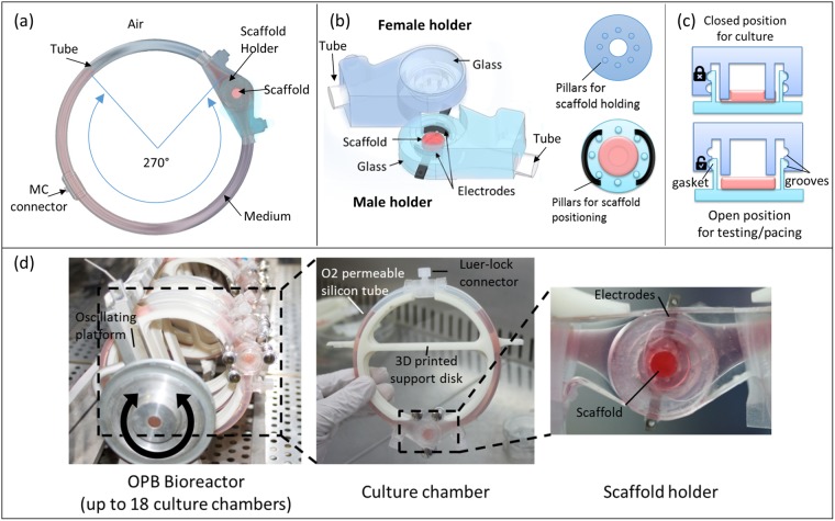
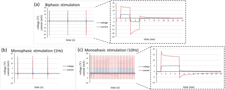
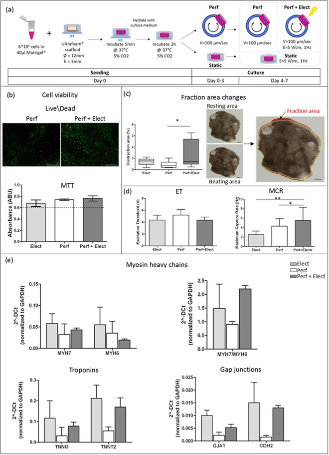
Tissue engineering strategies have been extensively exploited to generate functional cardiac patches. To maintain cardiac functionality in vitro, bioreactors have been designed to provide perfusion and electrical stimulation, alone or combined. However, due to several design limitations the integration of optical systems to assess cardiac maturation level is still missing within these platforms. Here we present a bioreactor culture chamber that provides 3D cardiac constructs with a bidirectional interstitial perfusion and biomimetic electrical stimulation, allowing direct cellular optical monitoring and contractility test. The chamber design was optimized through finite element models to house an innovative scaffold anchoring system to hold and to release it for the evaluation of tissue maturation and functionality by contractility tests. Neonatal rat cardiac fibroblasts subjected to a combined perfusion and electrical stimulation showed positive cell viability over time. Neonatal rat cardiomyocytes were successfully monitored for the entire culture period to assess their functionality. The combination of perfusion and electrical stimulation enhanced patch maturation, as evidenced by the higher contractility, the enhanced beating properties and the increased level of cardiac protein expression. This new multifunctional bioreactor provides a relevant biomimetic environment allowing for independently culturing, real-time monitoring and testing up to 18 separated patches.
Conflict of interest statement
The authors declare no competing interests.
Figures


Similar articles
-
Electric field stimulation integrated into perfusion bioreactor for cardiac tissue engineering.Tissue Eng Part C Methods. 2010 Dec;16(6):1417-26. doi: 10.1089/ten.TEC.2010.0068. Epub 2010 May 10. Tissue Eng Part C Methods. 2010. PMID: 20367291
-
Biomimetic perfusion and electrical stimulation applied in concert improved the assembly of engineered cardiac tissue.J Tissue Eng Regen Med. 2012 Nov;6(10):e12-23. doi: 10.1002/term.525. Epub 2011 Dec 13. J Tissue Eng Regen Med. 2012. PMID: 22170772 Free PMC article.
-
Electrical and mechanical stimulation of cardiac cells and tissue constructs.Adv Drug Deliv Rev. 2016 Jan 15;96:135-55. doi: 10.1016/j.addr.2015.07.009. Epub 2015 Jul 30. Adv Drug Deliv Rev. 2016. PMID: 26232525 Free PMC article. Review.
-
Myocardial scaffold-based cardiac tissue engineering: application of coordinated mechanical and electrical stimulations.Langmuir. 2013 Sep 3;29(35):11109-17. doi: 10.1021/la401702w. Epub 2013 Aug 20. Langmuir. 2013. PMID: 23923967 Free PMC article.
-
Analysis of the role of perfusion, mechanical, and electrical stimulation in bioreactors for cardiac tissue engineering.Bioprocess Biosyst Eng. 2024 Jun;47(6):767-839. doi: 10.1007/s00449-024-03004-5. Epub 2024 Apr 20. Bioprocess Biosyst Eng. 2024. PMID: 38643271 Review.
Cited by
-
Tissue Engineering Techniques for Induced Pluripotent Stem Cell Derived Three-Dimensional Cardiac Constructs.Tissue Eng Part B Rev. 2022 Aug;28(4):891-911. doi: 10.1089/ten.TEB.2021.0088. Epub 2021 Nov 23. Tissue Eng Part B Rev. 2022. PMID: 34476988 Free PMC article. Review.
-
Electroconductive Hydrogels for Tissue Engineering: Current Status and Future Perspectives.Bioelectricity. 2020 Sep 1;2(3):279-292. doi: 10.1089/bioe.2020.0025. Epub 2020 Sep 16. Bioelectricity. 2020. PMID: 34476358 Free PMC article.
-
Role of Cav1.3 Channels in Brain-Heart Interactions: An Unexpected Journey.Biomedicines. 2025 Jun 4;13(6):1376. doi: 10.3390/biomedicines13061376. Biomedicines. 2025. PMID: 40564095 Free PMC article. Review.
-
Insights to Heart Development and Cardiac Disease Models Using Pluripotent Stem Cell Derived 3D Organoids.Front Cell Dev Biol. 2021 Dec 2;9:788955. doi: 10.3389/fcell.2021.788955. eCollection 2021. Front Cell Dev Biol. 2021. PMID: 34926467 Free PMC article. Review.
-
Two Decades of Advances and Limitations in Organ Recellularization.Curr Issues Mol Biol. 2024 Aug 22;46(8):9179-9214. doi: 10.3390/cimb46080543. Curr Issues Mol Biol. 2024. PMID: 39194760 Free PMC article. Review.
References
-
- Mozaffarian D, et al. Heart disease and stroke statistics-2016 update a report from the American Heart Association. Circulation. 2016;133:e38–e48. - PubMed
Publication types
MeSH terms
Substances
LinkOut - more resources
Full Text Sources
Other Literature Sources
