. 2018 Nov 16;8(1):16958.
doi: 10.1038/s41598-018-35287-6.

Cued reactivation during slow-wave sleep induces brain connectivity changes related to memory stabilization

Affiliations

Cued reactivation during slow-wave sleep induces brain connectivity changes related to memory stabilization

Ruud M W J Berkers et al. Sci Rep. .

Abstract

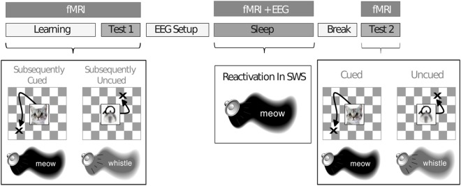
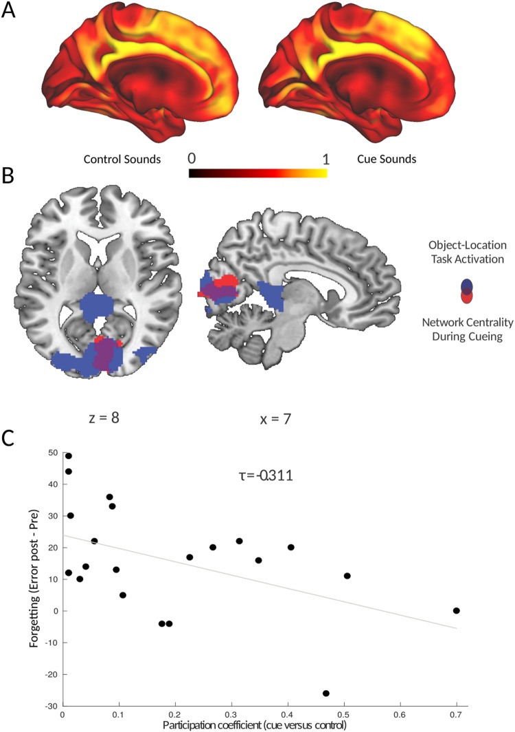
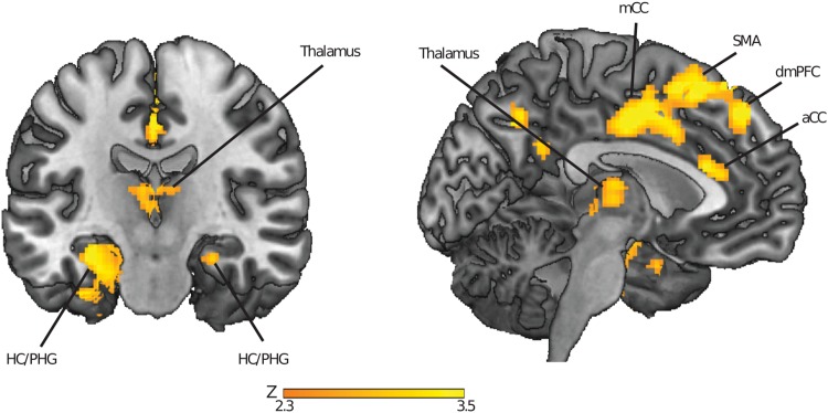
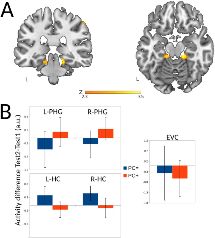
Memory reprocessing following acquisition enhances memory consolidation. Specifically, neural activity during encoding is thought to be 'replayed' during subsequent slow-wave sleep. Such memory replay is thought to contribute to the functional reorganization of neural memory traces. In particular, memory replay may facilitate the exchange of information across brain regions by inducing a reconfiguration of connectivity across the brain. Memory reactivation can be induced by external cues through a procedure known as "targeted memory reactivation". Here, we analysed data from a published study with auditory cues used to reactivate visual object-location memories during slow-wave sleep. We characterized effects of memory reactivation on brain network connectivity using graph-theory. We found that cue presentation during slow-wave sleep increased global network integration of occipital cortex, a visual region that was also active during retrieval of object locations. Although cueing did not have an overall beneficial effect on the retention of cued versus uncued associations, individual differences in overnight memory stabilization were related to enhanced network integration of occipital cortex. Furthermore, occipital cortex displayed enhanced connectivity with mnemonic regions, namely the hippocampus, parahippocampal gyrus, thalamus and medial prefrontal cortex during cue sound presentation. Together, these results suggest a neural mechanism where cue-induced replay during sleep increases integration of task-relevant perceptual regions with mnemonic regions. This cross-regional integration may be instrumental for the consolidation and long-term storage of enduring memories.


Conflict of interest statement

The authors declare no competing interests.

Figures

Figure 1
Figure 1
Schematic depiction of the experimental procedure. Participants learned 50 object-location associations, presented simultaneously with object-related sounds, inside the MR-scanner. After a baseline test (Test 1), participants were set up for polysomnographic recordings and went to sleep inside the MR-scanner. Half of the learned associations were cued with sounds during slow-wave sleep. After awaking and a short break, participants performed another post-sleep test (Test 2). Reprinted from.
Figure 2
Figure 2
Reactivation-related changes in network integration predict memory stabilization. (A) Participation Coefficient mapped for every voxel in the brain during the presentation of control sounds (not previously paired with object-location information, left panel) and cue sounds (previously paired with object-location associations, right panel). (B) Increased network integration was found only in the occipital cortex during the presentation of cue sounds versus control sounds during slow-wave sleep. This region overlapped with the set of regions activated in response to the object-location association task. Parametric maps were superimposed onto a template brain, using the cluster-forming threshold of Z > 2.33. (C) The increase in participation coefficient of the occipital cortex during cueing predicted memory stabilization, indicated by reduced forgetting (expressed as the difference in error distances) between test 1 (pre) and test 2 (post).
Figure 3
Figure 3
Regions displaying greater coupling with occipital cortex during cueing in slow-wave sleep. Parametric maps were superimposed onto a template brain, using the cluster-forming threshold of Z > 2.33. HC = Hippocampus, PHG = Parahippocampal Gyrus, aCC = Anterior Cingulate Cortex, mCC = Middle Cingulate Cortex, SMA = Supplementary Motor Area, dmPFC = Dorsomedial Prefrontal Cortex.
Figure 4
Figure 4
Overnight activity changes related to network integration of occipital cortex (A) Regions in the parahippocampal gyrus where there is a relation, on the one hand, between an activity increase between the pre-sleep and post-sleep test and, on the other hand, increased network integration of occipital cortex during cueing in the between Test1 and Test 2. Parametric maps were superimposed onto a template brain, using the cluster-forming threshold of Z > 2.33. (B) For comparison purposes, the mean overnight regional activation differences (calculated as activity during post-sleep Test 2 minus pre-sleep Test 1) are plotted for the left (L-PHG) and right (R-PHG) parahippocampal gyrus, the left (L-HC) and right (R-HC) hippocampus, and early visual cortex (EVC) for two groups of participants based on a median split on the scores of the participation coefficient in the early visual cortex. Even though there were no significant differences here, the numerical values are broadly in agreement with our hypotheses, namely finding a reduced activity of the hippocampus at Test 2 and an increased activity of the parahippocampal gyrus for the PC+ group (those participants that did display the strongest increase of the participation coefficient) versus the PC = group (those participants that displayed the lowest increase of the participation coefficient)and no visible differences in the early visual cortex.

References

    1. Diekelmann S, Büchel C, Born J, Rasch B. Labile or stable: opposing consequences for memory when reactivated during waking and sleep. Nat. Neurosci. 2011;14:381–386. doi: 10.1038/nn.2744. - DOI - PubMed
    1. Diekelmann S, Born J. The memory function of sleep. Nat. Rev. Neurosci. 2010;11:114–126. doi: 10.1038/nrn2762. - DOI - PubMed
    1. Wilson MA, McNaughton BL. Reactivation of hippocampal ensemble memories during sleep. Science. 1994;265:676–679. doi: 10.1126/science.8036517. - DOI - PubMed
    1. Ji D, Wilson MA. Coordinated memory replay in the visual cortex and hippocampus during sleep. Nat. Neurosci. 2007;10:100–107. doi: 10.1038/nn1825. - DOI - PubMed
    1. Euston DR, Tatsuno M, McNaughton BL. Fast-forward playback of recent memory sequences in prefrontal cortex during sleep. Science. 2007;318:1147–1150. doi: 10.1126/science.1148979. - DOI - PubMed

Publication types