

Enhanced protection conferred by mucosal BCG vaccination associates with presence of antigen-specific lung tissue-resident PD-1+ KLRG1- CD4+ T cells
- PMID: 30446726
- PMCID: PMC7051908
- DOI: 10.1038/s41385-018-0109-1
Enhanced protection conferred by mucosal BCG vaccination associates with presence of antigen-specific lung tissue-resident PD-1+ KLRG1- CD4+ T cells
Abstract
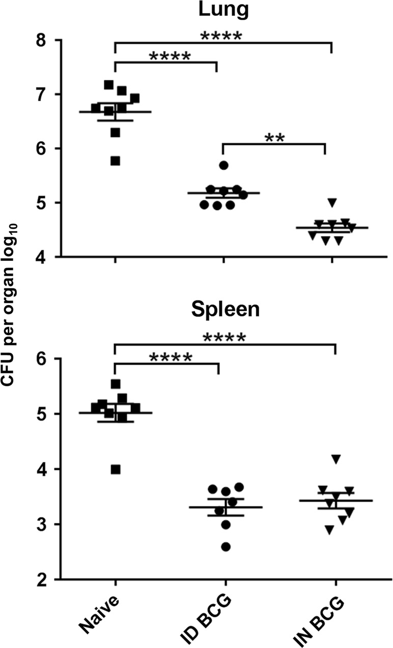
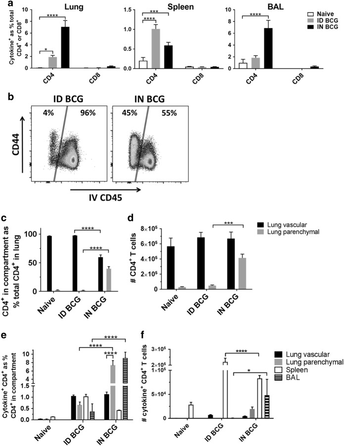
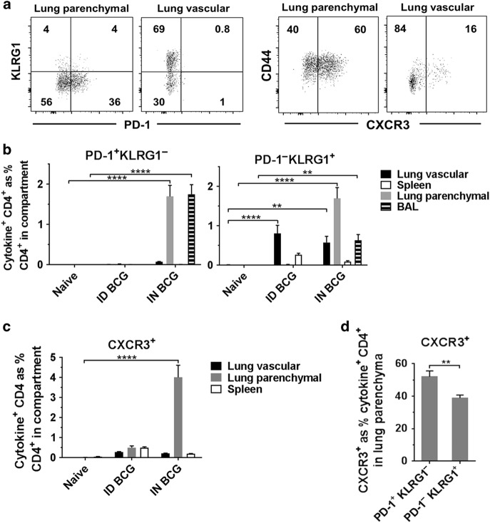
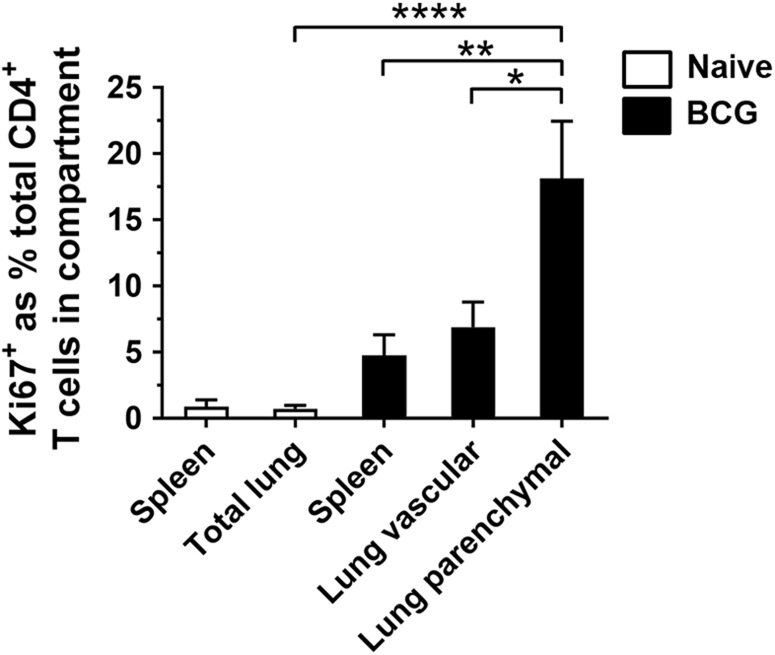
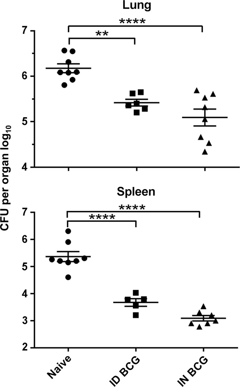
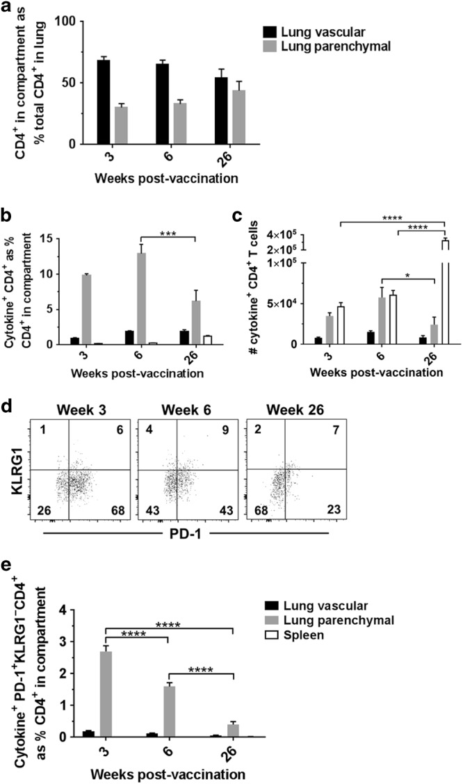
BCG, the only vaccine licensed against tuberculosis, demonstrates variable efficacy in humans. Recent preclinical studies highlight the potential for mucosal BCG vaccination to improve protection. Lung tissue-resident memory T cells reside within the parenchyma, potentially playing an important role in protective immunity to tuberculosis. We hypothesised that mucosal BCG vaccination may enhance generation of lung tissue-resident T cells, affording improved protection against Mycobacterium tuberculosis. In a mouse model, mucosal intranasal (IN) BCG vaccination conferred superior protection in the lungs compared to the systemic intradermal (ID) route. Intravascular staining allowed discrimination of lung tissue-resident CD4+ T cells from those in the lung vasculature, revealing that mucosal vaccination resulted in an increased frequency of antigen-specific tissue-resident CD4+ T cells compared to systemic vaccination. Tissue-resident CD4+ T cells induced by mucosal BCG displayed enhanced proliferative capacity compared to lung vascular and splenic CD4+ T cells. Only mucosal BCG induced antigen-specific tissue-resident T cells expressing a PD-1+ KLRG1- cell-surface phenotype. These cells constitute a BCG-induced population which may be responsible for the enhanced protection observed with IN vaccination. We demonstrate that mucosal BCG vaccination significantly improves protection over systemic BCG and this correlates with a novel population of BCG-induced lung tissue-resident CD4+ T cells.
Conflict of interest statement
The authors declare no competing interests.
Figures
Similar articles
-
Mucosal BCG Vaccination Induces Protective Lung-Resident Memory T Cell Populations against Tuberculosis.mBio. 2016 Nov 22;7(6):e01686-16. doi: 10.1128/mBio.01686-16. mBio. 2016. PMID: 27879332 Free PMC article.
-
Systemic BCG immunization induces persistent lung mucosal multifunctional CD4 T(EM) cells which expand following virulent mycobacterial challenge.PLoS One. 2011;6(6):e21566. doi: 10.1371/journal.pone.0021566. Epub 2011 Jun 24. PLoS One. 2011. PMID: 21720558 Free PMC article.
-
Attrition of T-cell functions and simultaneous upregulation of inhibitory markers correspond with the waning of BCG-induced protection against tuberculosis in mice.PLoS One. 2014 Nov 24;9(11):e113951. doi: 10.1371/journal.pone.0113951. eCollection 2014. PLoS One. 2014. PMID: 25419982 Free PMC article.
-
[Novel vaccines against M. tuberculosis].Kekkaku. 2006 Dec;81(12):745-51. Kekkaku. 2006. PMID: 17240920 Review. Japanese.
-
BCG - old workhorse, new skills.Curr Opin Immunol. 2017 Aug;47:8-16. doi: 10.1016/j.coi.2017.06.007. Epub 2017 Jul 15. Curr Opin Immunol. 2017. PMID: 28719821 Review.
Cited by
-
Phenotypic and Immunometabolic Aspects on Stem Cell Memory and Resident Memory CD8+ T Cells.Front Immunol. 2022 Jun 17;13:884148. doi: 10.3389/fimmu.2022.884148. eCollection 2022. Front Immunol. 2022. PMID: 35784300 Free PMC article. Review.
-
Infection resisters: targets of new research for uncovering natural protective immunity against Mycobacterium tuberculosis.F1000Res. 2019 Sep 27;8:F1000 Faculty Rev-1698. doi: 10.12688/f1000research.19805.1. eCollection 2019. F1000Res. 2019. PMID: 31602294 Free PMC article. Review.
-
The Yin and Yang of Targeting KLRG1+ Tregs and Effector Cells.Front Immunol. 2022 Apr 29;13:894508. doi: 10.3389/fimmu.2022.894508. eCollection 2022. Front Immunol. 2022. PMID: 35572605 Free PMC article. Review.
-
The Immunogenicity and Safety of Mycobacterium tuberculosis-mosR-Based Double Deletion Strain in Mice.Microorganisms. 2023 Aug 18;11(8):2105. doi: 10.3390/microorganisms11082105. Microorganisms. 2023. PMID: 37630665 Free PMC article.
-
BCG vaccination strategies against tuberculosis: updates and perspectives.Hum Vaccin Immunother. 2021 Dec 2;17(12):5284-5295. doi: 10.1080/21645515.2021.2007711. Epub 2021 Dec 2. Hum Vaccin Immunother. 2021. PMID: 34856853 Free PMC article. Review.
References
-
- Global Tuberculosis Report (WHO, Geneva, 2015).
Publication types
MeSH terms
Substances
Grants and funding
LinkOut - more resources
Full Text Sources
Other Literature Sources
Medical
Research Materials
