Review
. 2019 Dec;22(4):503-512.
doi: 10.1007/s40477-018-0341-2. Epub 2018 Nov 16.

Ultrasonography of the pediatric spleen: a pictorial essay

Affiliations
Review

Ultrasonography of the pediatric spleen: a pictorial essay

Marco Di Serafino et al. J Ultrasound. 2019 Dec.

Abstract

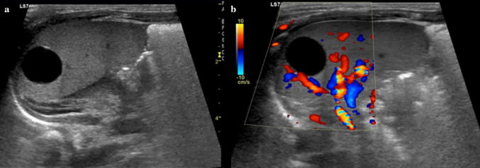
In infants and children, the spleen is involved in many pathological processes, whether those processes are isolated or related to systemic diseases. Pathology of the pediatric spleen includes congenital anomalies, splenomegaly, trauma, focal lesions, infarction, and tumors. Ultrasonography (US) is a widely available, fast, noninvasive imaging technique to assess the size, shape, and position of the spleen, as well as to define splenic echotexture. US is capable of screening for splenic disorders without the risk of ionizing radiation; it is the initial imaging examination performed to evaluate suspected splenic pathology, providing clinicians with helpful decisional support. US plays an important role in the detection of even very small amounts of hemoperitoneum, a herald of significant abdominal organ injury, in pediatric blunt abdominal trauma. Moreover, contrast-enhanced US may allow early detection of splenic injuries, ideally minimizing children's risk from radiation exposure. This pictorial essay illustrates the normal ultrasound appearance of the pediatric spleen and the sonographic findings which may guide clinicians to a correct diagnosis of pathologic conditions.

In età pediatrica la milza è coinvolta in molti processi patologici, che possono manifestarsi come entità isolate o possono essere secondarie a malattie sistemiche. I quadri patologici splenici includono anomalie congenite, splenomegalia, trauma, lesioni focali, infarti e tumori. L’ecografia è una metodica largamente disponibile, veloce e non invasiva che permette di determinare le dimensioni, la posizione e la morfologia della milza e di caratterizzarne inoltre l’ecotessitura parenchimale. L’esame ecografico permette di identificare precocemente le alterazioni spleniche senza il rischio di radiazioni ionizzanti e viene per questo motivo utilizzato come metodica di imaging iniziale per valutare quadri sospetti di patologia splenica fornendo ai clinici un utile supporto decisionale. Nei traumi addominali chiusi, l’ecografia svolge un ruolo importante nell’identificare quantità persino esigue di emoperitoneo, reperto associato a significative lesioni degli organi addominali. Inoltre, l’utilizzo dell’ecografia con mezzo di contrasto potrebbe permettere di identificare precocemente lesioni traumatiche della milza, e ridurre, idealmente, al minimo le radiazioni nei pazienti pediatrici. Questo pictorial essay illustra il normale aspetto ecografico della milza in età pediatrica ed i reperti ecografici che possono condurre alla corretta diagnosi di processi patologici.

Keywords: Congenital anomalies; Focal splenic lesions; Pediatric sonography; Splenomegaly; Trauma; Tumors.


Conflict of interest statement

The authors declare that they have no conflict of interest.

Figures

Fig. 1
Fig. 1
Normal splenic appearances at transducers of different frequencies. Gray-scale images acquired with: a a convex array at 5.0 MHz b a linear array in trapezoidal format at 7.5 MHz c a linear array at 9.0 MHz and d at 14.0 MHz
Fig. 2
Fig. 2
The zebra pattern of splenic parenchyma. Longitudinal sonograms of spleens from different patients, obtained with curved-array transducer, show bands of hypoechogenicity (white arrows)
Fig. 3
Fig. 3
Accessory spleens. Coronal oblique ultrasound images, from different patients, show small ovoid nodules (white arrows) a, b near the splenic hilum and c, d at the lower splenic pole
Fig. 4
Fig. 4
Hypertrophied accessory spleen a Gray scale and b color Doppler images show a compensatory hypertrophy of an accessory spleen secondary to a previous splenectomy
Fig. 5
Fig. 5
Situs inversus with polysplenia. a Longitudinal sonogram of the right upper quadrant showing multiple right-sided splenic masses (white arrows). b Transverse ultrasound of the left upper quadrant demonstrating a left-sided liver. c Axial T2-weighted magnetic resonance image shows situs inversus with multiple splenic masses in the right upper quadrant (white arrows)
Fig. 6
Fig. 6
Splenic lobulation. Longitudinal ultrasound images, from different patients, show a, b lobulations along the medial part of the spleen and c two clefts at the lower splenic pole (white arrows)
Fig. 7
Fig. 7
Splenomegaly. Coronal oblique ultrasound image demonstrating the sonographic measurement of longitudinal and transverse dimensions of an enlarged spleen
Fig. 8
Fig. 8
Splenic trauma. a Longitudinal sonogram of the spleen following blunt trauma demonstrates an intraparenchymal laceration that presents with a linear hypoechoic area (white arrow). b Axial computed tomography after contrast confirms the splenic lesion (white arrow) and shows a left hepatic lobe laceration (black-headed arrow)
Fig. 9
Fig. 9
Splenic trauma. a Gray scale and b e-flow technique images show a mild heterogeneity of the splenic lower pole (thin white arrows) associated with the c presence of splenic capsular rupture (thick white arrows) and resultant perisplenic hematoma
Fig. 10
Fig. 10
CEUS in splenic trauma. a Longitudinal sonogram following blunt trauma demonstrates an area of ill-defined altered echogenicity at the middle of the spleen. b CEUS clearly demonstrates a hypoechoic lesion extending at the surface, suggestive of splenic laceration
Fig. 11
Fig. 11
Splenic infarction. a Longitudinal gray scale and b color Doppler images of the spleen showing a subcapsular hypoechoic segmental lesion, which is clearly demonstrated b as an area of absent color flow consistent with a splenic infarction
Fig. 12
Fig. 12
Wandering spleen. Ultrasound images of the spleen obtained in the right lateral decubitus position. a Longitudinal and b, c transverse views of the lower abdomen and pelvis demonstrating the displaced enlarged spleen in the pelvis, extending behind the bladder. d Interrogation with the e-flow technique shows normal blood flow within the enlarged spleen
Fig. 13
Fig. 13
Splenic cyst in a neonate. a Gray scale and b color Doppler images of the spleen show a round, sharply marginated anechoic cyst with no internal flow

Similar articles

Cited by

References

    1. Kahramaner Z, Erdemir A, Arik B, Bilgili G, Tekin M, Genc Y. Reference ranges of liver and spleen dimensions in term infants: sonographic measurements. J Med Ultrason (2001) 2015;42(1):77–81. doi: 10.1007/s10396-014-0578-0. - DOI - PubMed
    1. Rosenberg HK, Markowitz RI, Kolberg H, Park C, Hubbard A, Bellah RD. Normal splenic size in infants and children: sonographic measurements. AJR Am J Roentgenol. 1991;157(1):119–121. doi: 10.2214/ajr.157.1.2048509. - DOI - PubMed
    1. Megremis SD, Vlachonikolis IG, Tsilimigaki AM. Spleen length in childhood with US: normal values based on age, sex, and somatometric parameters. Radiology. 2004;231(1):129–134. doi: 10.1148/radiol.2311020963. - DOI - PubMed
    1. Safak AA, Simsek E, Bahcebasi T. Sonographic assessment of the normal limits and percentile curves of liver, spleen, and kidney dimensions in healthy school-aged children. J Ultrasound Med. 2005;24(10):1359–1364. doi: 10.7863/jum.2005.24.10.1359. - DOI - PubMed
    1. Konus OL, Ozdemir A, Akkaya A, Erbas G, Celik H, Isik S. Normal liver, spleen, and kidney dimensions in neonates, infants, and children: evaluation with sonography. AJR Am J Roentgenol. 1998;171(6):1693–1698. doi: 10.2214/ajr.171.6.9843315. - DOI - PubMed