Comparative Study
. 2018 Dec;109(12):3993-4002.
doi: 10.1111/cas.13806. Epub 2018 Nov 16.

Immunomodulatory activity of lenvatinib contributes to antitumor activity in the Hepa1-6 hepatocellular carcinoma model

Affiliations
Comparative Study

Immunomodulatory activity of lenvatinib contributes to antitumor activity in the Hepa1-6 hepatocellular carcinoma model

Takayuki Kimura et al. Cancer Sci. 2018 Dec.

Abstract

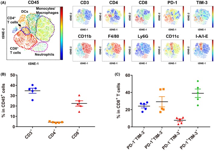
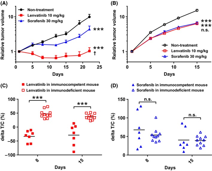
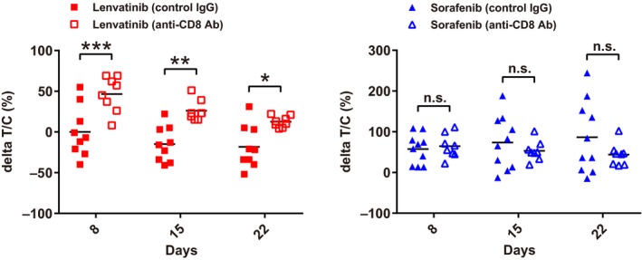
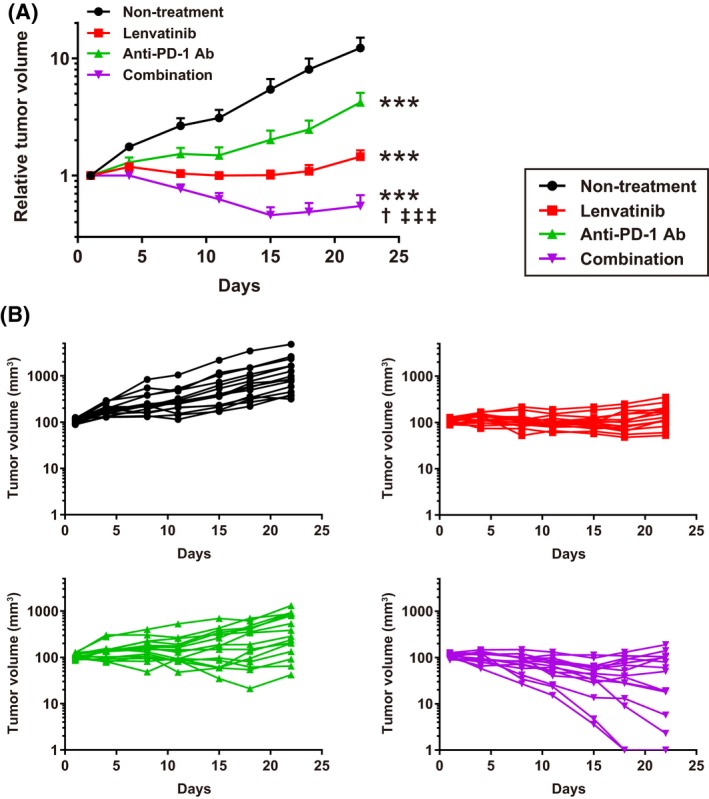
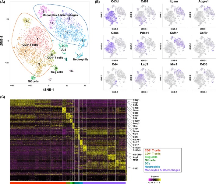
Angiogenesis inhibitors such as lenvatinib and sorafenib, and an immune checkpoint inhibitor (ICI), nivolumab, are used for anticancer therapies against advanced hepatocellular carcinoma (HCC). Combination treatments comprising angiogenesis inhibitors plus ICIs are promising options for improving clinical benefits in HCC patients, and clinical trials are ongoing. Here, we investigated the antitumor and immunomodulatory activities of lenvatinib (a multiple receptor tyrosine kinase inhibitor targeting vascular endothelial growth factor receptor 1-3, fibroblast growth factor receptor 1-4, platelet-derived growth factor receptor α, KIT and RET) and the combined antitumor activity of lenvatinib plus anti-programmed cell death 1 (PD-1) antibody in the Hepa1-6 mouse HCC syngeneic model. We found that the antitumor activities of lenvatinib and sorafenib were not different in immunodeficient mice, but lenvatinib showed more potent antitumor activity than sorafenib in immunocompetent mice. The antitumor activity of lenvatinib was greater in immunocompetent mice than in immunodeficient mice and was attenuated by CD8+ T cell depletion. Treatment with lenvatinib plus anti-PD-1 antibody resulted in more tumor regression and a higher response rate compared with either treatment alone in immunocompetent mice. Single-cell RNA sequencing analysis demonstrated that treatment with lenvatinib with or without anti-PD-1 antibody decreased the proportion of monocytes and macrophages population and increased that of CD8+ T cell populations. These data suggest that lenvatinib has immunomodulatory activity that contributes to the antitumor activity of lenvatinib and enhances the antitumor activity in combination treatment with anti-PD-1 antibody. Combination treatment of lenvatinib plus anti-PD-1 antibody therefore warrants further investigation against advanced HCC.

Keywords: anti-PD-1 antibody; hepatocellular carcinoma; immunomodulatory activity; lenvatinib; sorafenib.


Figures

Figure 1
Figure 1
Characterization of immune cell populations in the Hepa1‐6 tumor model in C57L/J mice. A, Tumor‐infiltrating lymphocytes from Hepa1‐6 syngeneic mice were examined by flow cytometry analysis followed by viSNE analysis. For each molecule examined, the expression level in individual cells is indicated by the right color bar (red: high, blue: low). Representative data are shown (n = 5). B, Percentages of CD3+, CD4+ or CD8+ T cells in CD45+ cells are shown. C, Percentages of PD‐1+/− TIM‐3+/− cells in CD8+ T cells are shown. Data are shown as means ± SEM. The data shown are representative of 2 independent experiments
Figure 2
Figure 2
Antitumor activities of lenvatinib and sorafenib in Hepa1‐6 mouse tumor models. Mice were orally administered 10 mg/kg of lenvatinib or 30 mg/kg of sorafenib once daily, or subjected to non‐treatment (control). The day on which treatment commenced was designated as day 1. Relative tumor volumes of (A) Hepa1‐6 tumor model in immunocompetent mice, C57L/J (n = 7) or (B) Hepa1‐6 tumor model in immunodeficient mice (n = 10). Data are shown as means + SEM. ***< .001 vs non‐treatment control; †< .05, n.s., not significant between lenvatinib and sorafenib (Dunnett's multiple comparisons test). C, Antitumor activities shown as ΔT/C values of lenvatinib and sorafenib in Hepa1‐6 mice tumor models at days 8 and 15. ***< .001, n.s., not significant between immunocompetent and immunodeficient mice (Sidak's multiple comparisons test). The data shown are representative of 2 independent experiments
Figure 3
Figure 3
Antitumor activities of lenvatinib and sorafenib in Hepa1‐6 tumor model in C57L/J mice with CD8+ T cell depletion. ΔT/C values at days 8, 15 and 22 (mouse isotype control IgG [control IgG], n = 9‐10; anti‐CD8α antibody [anti‐CD8 Ab], n = 8). ***< .001, **< .01, *< .05, n.s., not significant between control IgG and anti‐CD8 Ab treated mice (Sidak's multiple comparisons test). The data shown are representative of 2 independent experiments
Figure 4
Figure 4
Antitumor activities of lenvatinib, anti‐mouse PD‐1 antibody (anti‐PD‐1 Ab), and combination of lenvatinib plus anti‐PD‐1 Ab in Hepa1‐6 mouse tumor models. Mice were orally administered 10 mg/kg of lenvatinib once daily, intraperitoneally injected with 200 μg/head of anti‐PD‐1 Ab twice weekly, subjected to a combination of both treatments, or subjected to non‐treatment (control). A, Relative tumor volume at indicated time points (n = 15). Data are shown as means + SEM. ***< .001 vs non‐treatment control; †< .05 vs lenvatinib; ‡‡‡< .001 vs anti‐PD‐1 Ab (Dunnett's multiple comparisons test). B, Spider plots of each individual mouse's tumor volume in each group. Nonpalpable size tumors were defined as 1 mm3 tumor volume in the graph. The data shown are representative of 2 independent experiments
Figure 5
Figure 5
Immune cell population analysis in Hepa1‐6 syngeneic mouse tumors by single‐cell RNA sequencing. A, Two‐dimensional tSNE plot depicting 7456 single cells, each classified into 1 of the 17 clusters shown with distinct colors. CD8+ T cells, and monocytes and macrophages were surrounded by orange and blue broken lines, respectively. B, tSNE projection for 12 representative cell markers used for defining cell types of clusters. Each cell is colored by expression of the marker gene, with deep purple indicating high normalized unique molecular identifier. C, Heatmap of scaled gene expressions of the top 10 cluster‐specific genes detected from each cluster. Each row represents 1 gene and each column shows 1 cell ordered by cluster number

Similar articles

Cited by

References

    1. Llovet JM, Zucman‐Rossi J, Pikarsky E, et al. Hepatocellular carcinoma. Nat Rev Dis Primers. 2016;2:16018. - PubMed
    1. McGlynn KA, Petrick JL, London WT. Global epidemiology of hepatocellular carcinoma: an emphasis on demographic and regional variability. Clin Liver Dis. 2015;19:223‐238. - PMC - PubMed
    1. Torre LA, Bray F, Siegel RL, Ferlay J, Lortet‐Tieulent J, Jemal A. Global cancer statistics, 2012. CA Cancer J Clin. 2015;65:87‐108. - PubMed
    1. Wong MC, Jiang JY, Goggins WB, et al. International incidence and mortality trends of liver cancer: a global profile. Sci Rep. 2017;7:45846. - PMC - PubMed
    1. Llovet JM, Ricci S, Mazzaferro V, et al. Sorafenib in advanced hepatocellular carcinoma. N Engl J Med. 2008;359:378‐390. - PubMed

Publication types

MeSH terms