. 2018 Nov 17;15(1):321.
doi: 10.1186/s12974-018-1361-8.

Anti-inflammatory effects induced by pharmaceutical substances on inflammatory active brain astrocytes-promising treatment of neuroinflammation

Affiliations

Anti-inflammatory effects induced by pharmaceutical substances on inflammatory active brain astrocytes-promising treatment of neuroinflammation

Elisabeth Hansson et al. J Neuroinflammation. .

Abstract

Background: Pharmaceutical treatment with probable anti-inflammatory substances that attack cells in various ways including receptors, ion channels, or transporter systems may slow down the progression of inflammatory conditions. Astrocytes and microglia are the most prominent target cells for inflammation in the central nervous system. Their responses upon inflammatory stimuli work through the NO/cyclic GMP/protein kinase G systems that can downregulate the ATP-induced Ca2+ signaling, as well as G protein activities which alter Na+ transporters including Na+/K+-ATPase pump activity, Toll-like receptor 4 (TLR4), glutamate-induced Ca2+ signaling, and release of pro-inflammatory cytokines. The rationale for this project was to investigate a combination of pharmaceutical substances influencing the NO and the Gi/Gs activations of inflammatory reactive cells in order to make the cells return into a more physiological state. The ATP-evoked Ca2+ signaling is important maybe due to increased ATP release and subsequent activation of purinergic receptors. A balance between intercellular Ca2+ signaling through gap junctions and extracellular signaling mediated by extracellular ATP may be important for physiological function.

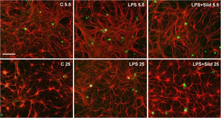
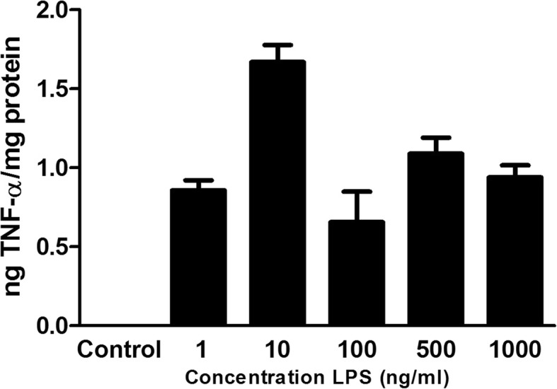
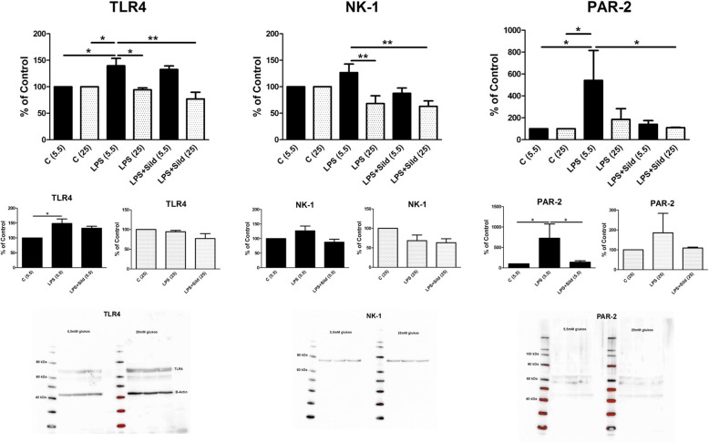
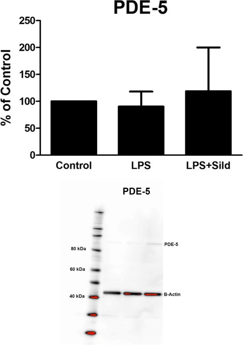
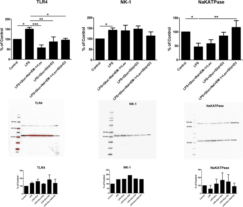
Methods: Astrocytes in primary cultures were incubated with lipopolysaccharide in a physiological glucose concentration for 24 h to induce inflammatory reactivity. The probable anti-inflammatory substances sildenafil and 1α,25-Dihydroxyvitamin D3 together with endomorphin-1, naloxone, and levetiracetam, were used in the presence of high glucose concentration in the medium to restore the cells. Glutamate-, 5-HT-, and ATP-evoked intracellular Ca2+ release, Na+/K+-ATPase expression, expression of inflammatory receptors, and release of tumor necrosis factor alpha were measured.

Results: Sildenafil in ultralow concentration together with 1α,25-Dihydroxyvitamin D3 showed most prominent effects on the ATP-evoked intracellular Ca2+ release. The μ-opioid agonist endomorphin-1, the μ-opioid antagonist naloxone in ultralow concentration, and the antiepileptic agent levetiracetam downregulated the glutamate-evoked intracellular Ca2+ release and TLR4. The combination of the pharmaceutical substances in high glucose concentration downregulated the glutamate- and ATP-evoked Ca2+ signaling and the TLR4 expression and upregulated the Na+/K+-ATPase pump.

Conclusion: Pharmaceutical treatment with the combination of substances that have potential anti-inflammatory effects, which attack different biochemical mechanisms in the cells may exert decisive effects to downregulate neuroinflammation in the nervous system.

Keywords: Astrocytes; Ca2+ signaling; Gap junction-coupled cells; Glucose; Inflammation; Na+/K+-ATPase; Pharmaceuticals; Restoration; TLR4.


Conflict of interest statement

Competing interest

The authors declare that they have no competing interests.

Ethics approval and consent to participate

We have bought the cell cultures from 3H Biomedical Science, Uppsala, Sweden, and used the cultures according to the manufacturer’s instructions. Therefore, no ethics approval can be done.

Consent for publication

Not applicable

Publisher’s Note

Springer Nature remains neutral with regard to jurisdictional claims in published maps and institutional affiliations.

Figures

Fig. 1
Fig. 1
Culture stained for OX42, a marker for microglial cells (green), and GFAP, a marker for astrocytes (red). Some microglia, less than 5%, were found in the control cultures ©, 5.5 mM glucose. The amount of microglia increased to 5–10% after treatment with LPS for 24 h in 5.5 mM glucose. The amount of microglia decreased after treatment with LPS for 24 h followed by incubation with LPS and sildenafil (Sild) for another 24 h in 5.5 mM glucose to less than 5%. Cultures cultivated in 25 mM glucose were less affected, 1–2%. Scale bar = 50 μm. Representative images are presented
Fig. 2
Fig. 2
LPS concentration curve verified by TNF-α release. n = 6
Fig. 3
Fig. 3
Astrocytes were stimulated, in a fluorescence-based assay for detecting changes in intracellular Ca2+ over time, with the following: 5-HT (10−5 M), glutamate (10−3 M), or ATP (10−4 M). The cells were cultivated in 5.5 mM or 25 mM glucose the whole cultivation period. Ca2+ responses when incubated with LPS (10 ng/ml) for 24 h, or when incubated with LPS for 24 h followed by LPS and sildenafil (Sild) (1 μM); unstimulated cells were used as controls ©. The area under the Ca2+ peak (AUC) was calculated for each Ca2+ transient, and the amplitude (peak) was expressed as the maximum increase. The cells were obtained from three experiments with quadruple wells in each. The level of significance was calculated against LPS (5.5) and analyzed using one-way ANOVA followed by Dunnett’s multiple comparisons test. *P < 0.05, **P < 0.01, ***P < 0.001. Separate small histograms show cells cultivated in 5.5 mM glucose and 25 mM glucose, respectively. The level of significance was calculated against LPS (5.5) or against LPS (25). *P < 0.05
Fig. 4
Fig. 4
Expression levels of the receptors TLR4, NK-1, and PAR-2 were studied using Western blot analysis. Astrocytes were cultivated in 5.5 mM or 25 mM glucose the whole cultivation period. The astrocytes were incubated with LPS (10 ng/ml) for 24 h or incubated with LPS for 24 h followed by LPS and sildenafil (Sild) (1 μM), or combination of all substances for another 24 h; unstimulated cells were used as controls ©. Statistical analysis: the level of significance was calculated against LPS (5.5) and analyzed using one-way ANOVA followed by Dunnett’s multiple comparisons test. *P < 0.05, **P < 0.01. n = 6. Separate small histograms show cells cultivated in 5.5 mM glucose and 25 mM glucose, respectively. The level of significance was calculated against LPS (5.5) or against LPS (25). *P < 0.05. Representative images of Western blot membranes are presented
Fig. 5
Fig. 5
The expression levels of PDE-5 were studied using Western blot analysis. Astrocytes were cultivated in 5.5 mM glucose the whole cultivation period. The astrocytes were incubated with LPS (10 ng/ml) for 24 h or incubated with LPS for 24 h followed by LPS and sildenafil (Sild) (1 μM) for another 24 h. n = 3. Representative image of Western blot membrane is presented
Fig. 6
Fig. 6
Astrocytes were stimulated, in a fluorescence-based assay for detecting changes in intracellular Ca2+ over time, with the following: 5-HT(10−5 M), glutamate (10−3 M), or ATP (10−4 M). AUC and peak values of Ca2+ transients are shown. The cells were cultivated in 5.5 mM glucose the whole cultivation period. Ca2+ responses were measured after the cells were incubated with LPS (10 ng/ml) for 24 h, when incubated with LPS for 24 h followed by LPS, 25 mM glucose, a combination of naloxone (Nal) (10−12 M), endomorphin-1 (EM-1) (10−6 M), and levetiracetam (Lev) (10−4 M), or a combination of sildenafil (Sild) (1 μM) and vitamin D3 (D3) (100 nM) or combination of all substances for another 24 h. Unstimulated cells were used as controls. The area under the Ca2+ peak (AUC) was calculated for each Ca2+ transient, and the amplitude (peak) was expressed as the maximum increase. The cells were obtained from four experiments with quadruple wells in each. The level of significance was calculated against LPS (5.5) and analyzed using one-way ANOVA followed by Dunnett’s multiple comparisons test. *P < 0.05, **P < 0.01, ***P < 0.001. Additional data, small histograms, for LPS + glucose, LPS + glucose + sildenafil, LPS + glucose + vitamin D3, and LPS + glucose + sildenafil + vitamin D3. * P < 0.05
Fig. 7
Fig. 7
Expression levels of TLR4, NK-1, and Na+/K+-ATPase were studied using Western blot analysis. Astrocytes were cultivated in 5.5 mM glucose the whole cultivation period. The cells were incubated with LPS (10 ng/ml) for 24 h, when incubated with LPS for 24 h followed by LPS, 25 mM glucose, a combination of naloxone (Nal) (10− 12 M), endomorphin-1 (EM-1) (10− 6 M), and levetiracetam (Lev) (10− 4 M), or a combination of sildenafil (Sild) (1 μM) and vitamin D3 (D3) (100 nM), or combination of all substances for another 24 h. Unstimulated cells were used as controls. The level of significance was calculated against LPS (5.5) and analyzed using one-way ANOVA followed by Dunnett’s multiple comparisons test. *P < 0.05, **P < 0.01, ***P < 0.001. n = 6. Representative images of Western blot membranes are presented. Additional data, small histograms below, show LPS + glucose, LPS + glucose + sildenafil, LPS + glucose + vitamin D3, and LPS + glucose + sildenafil + vitamin D3

References

    1. Perretti M, Leroy X, Bland EJ, Montero-Melendez T. Resolution pharmacology: opportunities for therapeutic innovation in inflammation. Trends Pharmacol Sci. 2015;36:737–755. doi: 10.1016/j.tips.2015.07.007. - DOI - PubMed
    1. Rönnbäck C, Hansson E. Gap junction coupled cells, barriers and systemic inflammation. Int J Open Access Ophthalmol. 2017;2(1):7. doi: 10.15226/2474-9249/2/1/00117. - DOI
    1. Peixoto CA, Gomes FOS. The role of phosphodiesterase-5 inhibitors in prostatic inflammation: a review. J Inflammation. 2015;12:54. doi: 10.1186/s12950-015-0099-7. - DOI - PMC - PubMed
    1. Cotrina ML, Lin JH, Alves-Rodrigues A, Liu S, Li J, Azmi-Ghadimi H, Kang J, Naus CC, Nedergaard M. Connexins regulate calcium signaling by controlling ATP release. Proc Natl Acad Sci U S A. 1998;95:15735–15740. doi: 10.1073/pnas.95.26.15735. - DOI - PMC - PubMed
    1. Liu X, Gangoso E, Yi C, Jeanson T, Kandelman S, Mantz J, Giaume C. General anesthetics have differential inhibitory effects on gap junction channels and hemichannels in astrocytes and neurons. GLIA. 2016;64:524–536. doi: 10.1002/glia.22946. - DOI - PubMed

MeSH terms