. 2018 Dec 4;115(11):2152-2166.
doi: 10.1016/j.bpj.2018.10.023. Epub 2018 Nov 2.

Dual Role of the C-Terminal Domain in Osmosensing by Bacterial Osmolyte Transporter ProP

Affiliations

Dual Role of the C-Terminal Domain in Osmosensing by Bacterial Osmolyte Transporter ProP

Doreen E Culham et al. Biophys J. .

Abstract

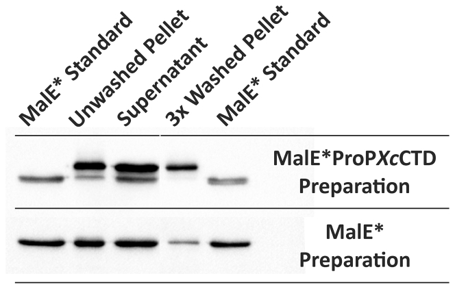
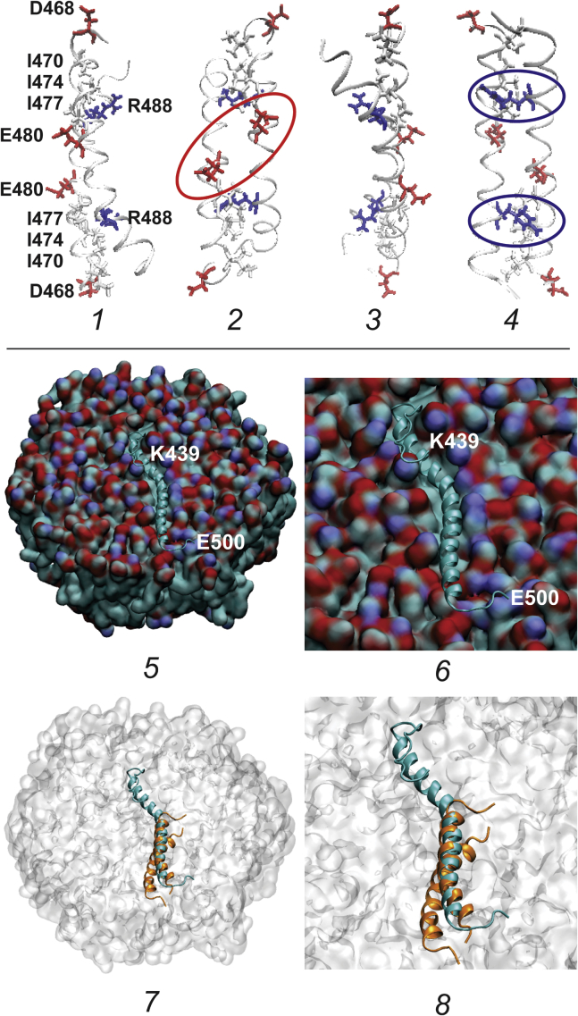
ProP is a member of the major facilitator superfamily, a proton-osmolyte symporter, and an osmosensing transporter. ProP proteins share extended cytoplasmic carboxyl terminal domains (CTDs) implicated in osmosensing. The CTDs of the best characterized, group A ProP orthologs, terminate in sequences that form intermolecular, antiparallel α-helical coiled coils (e.g., ProPEc, from Escherichia coli). Group B orthologs lack that feature (e.g., ProPXc, from Xanthomonas campestris). ProPXc was expressed and characterized in E. coli to further elucidate the role of the coiled coil in osmosensing. The activity of ProPXc was a sigmoid function of the osmolality in cells and proteoliposomes. ProPEc and ProPXc attained similar activities at the same expression level in E. coli. ProPEc transports proline and glycine betaine with comparable high affinities at low osmolality. In contrast, proline weakly inhibited high-affinity glycine-betaine uptake via ProPXc. The KM for proline uptake via ProPEc increases dramatically with the osmolality. The KM for glycine-betaine uptake via ProPXc did not. Thus, ProPXc is an osmosensing transporter, and the C-terminal coiled coil is not essential for osmosensing. The role of CTD-membrane interaction in osmosensing was examined further. As for ProPEc, the ProPXc CTD co-sedimented with liposomes comprising E. coli phospholipid. Molecular dynamics simulations illustrated association of the monomeric ProPEc CTD with the membrane surface. Comparison with the available NMR structure for the homodimeric coiled coil formed by the ProPEc-CTD suggested that membrane association and homodimeric coiled-coil formation by that peptide are mutually exclusive. The membrane fluidity in liposomes comprising E. coli phospholipid decreased with increasing osmolality in the range relevant for ProP activation. These data support the proposal that ProP activates as cellular dehydration increases cytoplasmic cation concentration, releasing the CTD from the membrane surface. For group A orthologs, this also favors α-helical coiled-coil formation that stabilizes the transporter in an active form.


Figures

Figure 1
Figure 1
Aligned sequences of the CTDs of ProP orthologs and paralogues. The sequences of the cytoplasmic CTDs of the ProP orthologs from E. coli (ProPEc), D. didantii (ProPDd, Q_47421), A. tumefaciens (ProPAt, NP_356328), X. campestris (ProPXc, NP_635478), and C. glutamicum (ProPCg, CAA_73136) are aligned with those of the closest ProP paralogues that are known not to be osmoregulatory transporters (shikimate transporter ShiA (12) (NP_416488) and α-ketoglutarate transporter KgtP (AAC_75640), both from E. coli). Residue 438 of ProPEc is intramembrane (64). Structural predictions: ag, coiled-coil heptads (81); bold, known coiled coil (44); H, α-helix; S, β-strand (82). Acidic residues are in red, and basic residues are in blue. To view this figure in color, go online.
Figure 2
Figure 2
ProPXc is a glycine-betaine transporter. E. coli strain WG350 pDM4 was cultivated in NaCl-free MOPS medium supplemented with L-arabinose at the indicated concentrations, and the initial rate of glycine-betaine uptake was measured as described in Materials and Methods. Error bars represent standard errors of the means for three replicate measurements. (A) Initial rates of glycine-betaine uptake were measured in unsupplemented MOPS medium with an osmolality of 0.15 mol/kg and at a glycine-betaine concentration of 100 μM. (B) MOPS medium was supplemented with L-arabinose (45 μM) and with NaCl to attain an osmolality of 0.24 mol/kg (open circles) or 0.45 mol/kg (closed circles). The regression lines were obtained by fitting the data to the Michaelis Menten equation by non-linear regression using Sigma-Plot 12.5. The resulting parameters are listed in Table 1.
Figure 3
Figure 3
Substrate specificities of the ProP orthologs. E. coli strains containing ProPXc (WG350 pDM4, A), ProPEc (WG350 pDC79, B), ProPCg (WG350 pYT12, C), and ProPAt (WG350 pYT13, D) were grown in NaCl-free MOPS medium. Expression of ProPAt was induced with 0.3 mM arabinose (36), and expression of ProPXc was induced with 0.4 mM arabinose. To optimize the activity of each transporter, the assay media were adjusted with NaCl to attain osmolalities of 0.15 mol/kg (A), 0.44 mol/kg (B and D), and 0.59 mol/kg (C). Assay media contained radiolabeled proline or glycine betaine at concentrations of 200 and 100 μM, respectively. Representative initial rates attained in assays without inhibitors were (nmoles/min/mg protein): 38 ± 3 (ProPXcA); 63 ± 1 (ProPEc, B); 47 ± 3 (ProPCg, C); and 59 ± 1 (ProPAt, D). Error bars represent standard errors of the means for three replicate measurements.
Figure 4
Figure 4
Osmolality dependence of osmolyte uptake via ProPXc and ProPEc in E. coli. E. coli strains WG350 pDC79 (open circles, ProPEc) and WG350 pDM4 (closed circles, ProPXc) were cultivated in NaCl-free MOPS medium that was unsupplemented (ProPEc) or supplemented with 0.4 mM arabinose (ProPXc). Initial rates of osmolyte uptake were measured as described in Materials and Methods using assay media adjusted to the indicated osmolalities with NaCl. Radiolabeled proline (for ProPEc) and glycine betaine (for ProPXc) were provided at concentrations of 200 and 100 μM, respectively. The arrow indicates the growth medium osmolality. Error bars represent standard errors of the means for three replicate measurements.
Figure 5
Figure 5
The absolute activities of His10-ProPXc, His10-ProPEc, and truncated His10-ProPEc variants in E. coli. E. coli WG350 derivatives harboring plasmids that encoded the indicated ProP variants were cultivated as for transport assays (see Materials and Methods). The media for the strains containing His10-ProPEc and His10-ProPXc were supplemented with D-glucose (2.2 mM) and L-arabinose (4 μM), respectively. (A–C) The transporter contents of cell extracts were compared by Western blotting with anti-ProPEc antibodies (21) (A) and anti-PentaHis antibodies (Qiagen, B). The loading of cell protein was determined by GelCode Blue (ThermoFisher Scientific) staining of a replicate sodium dodecyl sulfate polyacrylamide gel electrophoresis gel (C). ProPEc and His10-ProPEc were expressed at similar levels after no induction and D-glucose repression of the encoding genes, respectively (A). Further, densitometric analysis indicated that the expression levels of the other variants relative to His10-ProPEc (and hence ProPEc) were 0.42 for His10-ProPXc, 3.1 for His10-ProPEcΔ11, and 1.9 for His10-ProPEcΔ18 (B). (D) Initial rates of osmolyte uptake were measured as described in Materials and Methods using assay media adjusted to the indicated osmolalities with NaCl. Radiolabeled proline (for ProPEc and its variants) and glycine betaine (for ProPXc) were provided at concentrations of 200 and 100 μM, respectively. The activities of the His10-tagged transporters were corrected for their expression levels relative to ProPEc by applying the factors listed above. The symbols are ProPEc (closed circles), His10-ProPXc (open circles), His10-ProPEcΔ11 (closed inverted triangles), and His10-ProPEcΔ18 (closed triangles). Error bars represent standard errors of the means for three replicate measurements.
Figure 6
Figure 6
ProPXc is an osmosensing transporter. His10-ProPXc and ProPEc-His6 were purified and reconstituted in PRLs and initial rates of proline (ProPEc) or glycine-betaine (ProPXc) uptake were measured as a function of the assay medium osmolality as described in Materials and Methods. (A) Initial rates of osmolyte uptake are plotted as a function of the luminal K+ concentration, the latter calculated on the basis of the assay medium osmolality, the osmolality of the PRL preparation, and the assumption that PRLs behave as ideal osmometers (see inset) (16). The regression lines were obtained by fitting the data to Eq. 3 (16) and the regression parameters are summarized in (B) as means ± the asymptotic standard errors of the means.
Figure 7
Figure 7
The ProPXc CTD mediates the association of MalE with liposomes. Proteins MalEProPXcCTD and MalE were purified, and their association with liposomes was assessed with a sedimentation assay and Western blotting, employing anti-MalE as primary antibody, as described in Materials and Methods. The MalEProPXcCTD preparation included the full-length protein and a fragment that terminates at A449, whereas the MalE preparation included only the full-length protein (see the text, Fig. S2; Table S4). The gel lanes were loaded with purified MalE protein (as a marker), the pellet and supernatant obtained upon centrifugation of the initial peptide-liposome mixture, and the pellet obtained after washing the initial pellet three times.
Figure 8
Figure 8
Structure and membrane interaction of the ProPEc CTD. Representations of the structure of the ProPEc CTD as a homodimeric antiparallel coiled coil (14) and as a membrane-associated monomer (5 and 6) are shown. (14) Four representations of the NMR structure (PDB: 1R48) of the antiparallel, homodimeric α-helical coiled coil formed by peptides representing residues 468–497 of ProPEc (44) are shown. Each structure is rotated 90° around the vertical axis relative to the structure to the left. The backbones of the two peptides in each structure are colored gray, with the bonds of each N-terminal aspartate (residue 468) colored red. The bonds of isoleucine residues 470, 474, and 477 are colored gray; those of glutamate 480 are colored red; and those of arginine 488 are colored blue. In structure 2 (showing the “c/g” surface of the coiled coil), the glutamates 480 are circled in red. In structure 4 (showing the “b/e” surface of the coiled coil), the arginines 488 are circled in blue. (The c, g, b, and e refer to positions in the coiled-coil heptad repeat.) 5–8: The interaction of the entire, monomeric CTD of ProPEc (residues 439–500) with the surface of a membrane comprising PE (80%) and PG (20%) is shown. This structure was simulated as described in Materials and Methods. (5 and 6) The α-helical portion of the peptide (turquoise, residues 456–495) is embedded in the membrane, displacing the phospholipid headgroups. The figures represent a snapshot taken after 100 ns of simulation. (7 and 8) The NMR structure of the coiled-coil dimer (orange, residues 468–497) is superimposed on the simulated structure of the membrane-associated peptide monomer (turquoise) to show that coiled-coil formation and membrane association are mutually exclusive (see text).
Figure 9
Figure 9
Impact of the osmolality on membrane fluidity. The membrane fluidity was estimated by determining the fluorescence anisotropy (R) of DPH in liposomes prepared with a polar lipid extract from E. coli, as described in the text and Materials and Methods. Fluorescence anisotropies are reported as means ± the standard error of the mean for four replicate measurements. (A) DPH fluorescence anisotropy was measured as a function of temperature in 0.1 M potassium phosphate buffer with no added NaCl. (B) DPH fluorescence anisotropy was measured at 25°C as a function of the osmolality attained by adding NaCl (closed circles) or sorbitol (open circles) to the phosphate buffer. The sorbitol concentration was limited to avoid introducing viscosity effects. The luminal K+ concentration of the liposomes (see inset) was calculated on the basis that the phosphate buffer contained 0.178 M K+ and the liposomes behaved as ideal osmometers (42).
Figure 10
Figure 10
Proposed structural mechanism of osmosensing. ProPEc is believed to mediate proton-osmolyte symport via an alternating access mechanism in which its N- and C-terminal helix bundles can reorient, exposing the proton (H+) and osmolyte (gray circles) binding sites sequentially to the periplasm and cytoplasm. As an osmosensor, ProPEc may exist in three forms. In ProPI, the CTD associates with the membrane surface to lock the protein in an inactive, inward-facing conformation. In ProPA, the CTD is free and ProP is active, alternating between outward and inward-facing conformations. In ProPAC, the active species is stabilized as the CTDs form antiparallel, homodimeric coiled coils. Coiled-coil formation prevents the CTD from returning to the membrane surface because the arrangements of the CTDs in ProPI and ProPAC are mutually exclusive (Fig. 8). The distribution of ProPEc among these forms would be modulated by the osmolality: ProPI would be favored at low osmolality, whereas ProPA and ProPAC would be favored at increasingly high osmolality as the membrane fluidity decreases (see Fig. 9). ProP orthologs lacking the C-terminal coiled coil (e.g., ProPXc) would exist only as form ProPI or ProPA.

Similar articles

Cited by

References

    1. Sies M., Haussinger D. Osmosensing and Osmosignaling. Methods Enzymol. 2007;428:1–579. - PubMed
    1. Anishkin A., Loukin S.H., Kung C. Feeling the hidden mechanical forces in lipid bilayer is an original sense. Proc. Natl. Acad. Sci. USA. 2014;111:7898–7905. - PMC - PubMed
    1. Wood J.M. Bacterial responses to osmotic challenges. J. Gen. Physiol. 2015;145:381–388. - PMC - PubMed
    1. Wood J.M. Bacterial osmoregulation: a paradigm for the study of cellular homeostasis. Annu. Rev. Microbiol. 2011;65:215–238. - PubMed
    1. Murdock L., Burke T., Wood J.M. Analysis of strains lacking known osmolyte accumulation mechanisms reveals contributions of osmolytes and transporters to protection against abiotic stress. Appl. Environ. Microbiol. 2014;80:5366–5378. - PMC - PubMed

Publication types

MeSH terms

LinkOut - more resources