. 2018 Dec 4;115(11):2271-2277.
doi: 10.1016/j.bpj.2018.10.017. Epub 2018 Oct 30.

Bacterial Cohesion Predicts Spatial Distribution in the Larval Zebrafish Intestine

Affiliations

Bacterial Cohesion Predicts Spatial Distribution in the Larval Zebrafish Intestine

Brandon H Schlomann et al. Biophys J. .

Abstract

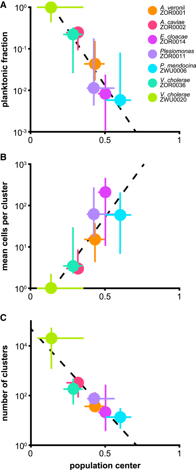
Are there general biophysical relationships governing the spatial organization of the gut microbiome? Despite growing realization that spatial structure is important for population stability, interbacterial competition, and host functions, it is unclear in any animal gut whether such structure is subject to predictive, unifying rules or if it results from contextual, species-specific behaviors. To explore this, we used light sheet fluorescence microscopy to conduct a high-resolution comparative study of bacterial distribution patterns throughout the entire intestinal volume of live, larval zebrafish. Fluorescently tagged strains of seven bacterial symbionts, representing six different species native to zebrafish, were each separately monoassociated with animals that had been raised initially germ-free. The strains showed large differences in both cohesion-the degree to which they auto-aggregate-and spatial distribution. We uncovered a striking correlation between each strain's mean position and its cohesion, whether quantified as the fraction of cells existing as planktonic individuals, the average aggregate size, or the total number of aggregates. Moreover, these correlations held within species as well; aggregates of different sizes localized as predicted from the pan-species observations. Together, our findings indicate that bacteria within the zebrafish intestine are subject to generic processes that organize populations by their cohesive properties. The likely drivers of this relationship-peristaltic fluid flow, tubular anatomy, and bacterial growth and aggregation kinetics-are common throughout animals. We therefore suggest that the framework introduced here of biophysical links between bacterial cohesion and spatial organization should be useful for directing explorations in other host-microbe systems, formulating detailed models that can quantitatively map onto experimental data, and developing new tools that manipulate cohesion to engineer microbiome function.


Figures

Figure 1
Figure 1
Diversity of bacterial population structures within the zebrafish intestine. (A) A schematic of a 5-day-old larval zebrafish. (B) A schematic of a larval zebrafish intestine with the three general anatomical regions and their approximate relative sizes highlighted. (C) Representative images from across the range of observed population structures. Each image is a maximal intensity projection of a full three-dimensional image stack, except for the top right inset, which is a single optical plane. Dashed amber lines trace the approximate boundaries of the intestine in each image. Examples of single cells (open arrowheads), small aggregates (closed arrowheads), and large aggregates (tailed arrowheads) are noted within insets under “subregion.” See also Videos S1, S2, S3, and S4. Top row: Populations of V. cholerae ZWU0020 localize to the anterior bulb and are dominated by highly motile planktonic cells (Video S1). Inset shows V. cholerae ZWU0020 cells in a different fish that was colonized with a 1:100 mixture of green and red variants. The dilute channel (green) is shown. Middle row: Populations of A. caviae ZOR0002 typically contain a range of bacterial aggregate sizes, as indicated by arrows. Inset shows a zoomed-in view of the same intestine. Bottom row: Populations of E. cloacae ZOR0014 typically consist of small numbers of large aggregates. Inset shows a zoomed-in view of the same intestine. To see this figure in color, go online.
Figure 2
Figure 2
Metrics of cohesion correlate with spatial distribution across bacterial strains. Shown are (A) the fraction of the population of each strain existing as single planktonic cells, (B) the average number of cells per cluster, and (C) the total number of clusters plotted against the population center, the center of mass position of each strain normalized by the length of the intestine. For the plots shown in (B) and (C), individual cells are considered clusters of size one. Circles show median values for each strain, and bars show 25 and 75% quartiles. Trendlines were generated from the unweighted linear regression of log10-transformed medians. To see this figure in color, go online.
Figure 3
Figure 3
Signatures of a cohesion-distribution relationship can be detected within populations at the strain level. For each strain shown, the size of every cluster with size two cells and greater across all samples is plotted against its normalized position along the intestine (small circles). Trendlines depict linear regressions of log10-transformed cluster sizes against position (black dashed lines). To better highlight trends, data were binned by position, and the mean SDs of cluster sizes were overlaid on each plot as large circles and bars. To see this figure in color, go online.

References

    1. Cox L.M., Yamanishi S., Blaser M.J. Altering the intestinal microbiota during a critical developmental window has lasting metabolic consequences. Cell. 2014;158:705–721. - PMC - PubMed
    1. Kundu P., Blacher E., Pettersson S. Our gut microbiome: the evolving inner self. Cell. 2017;171:1481–1493. - PubMed
    1. McKenney P.T., Pamer E.G. From hype to hope: the gut microbiota in enteric infectious disease. Cell. 2015;163:1326–1332. - PMC - PubMed
    1. Mark Welch J.L., Hasegawa Y., Borisy G.G. Spatial organization of a model 15-member human gut microbiota established in gnotobiotic mice. Proc. Natl. Acad. Sci. USA. 2017;114:E9105–E9114. - PMC - PubMed
    1. Tropini C., Earle K.A., Sonnenburg J.L. The gut microbiome: connecting spatial organization to function. Cell Host Microbe. 2017;21:433–442. - PMC - PubMed

Publication types

LinkOut - more resources