. 2019 Feb;189(2):283-294.
doi: 10.1016/j.ajpath.2018.10.012. Epub 2018 Nov 15.

Inflammatory Stress Causes N-Glycan Processing Deficiency in Ocular Autoimmune Disease

Affiliations

Inflammatory Stress Causes N-Glycan Processing Deficiency in Ocular Autoimmune Disease

Ashley M Woodward et al. Am J Pathol. 2019 Feb.

Abstract

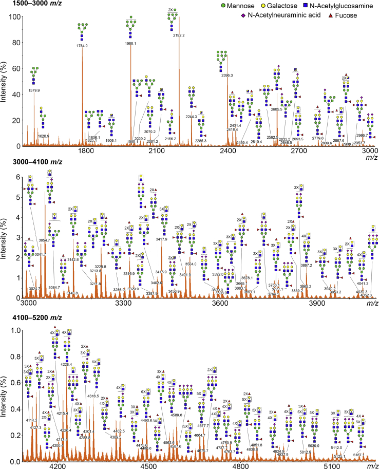
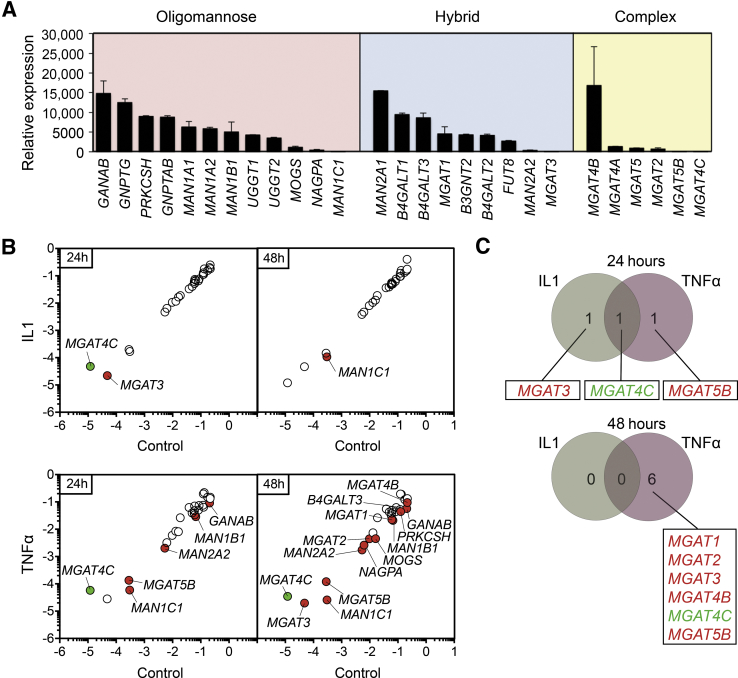
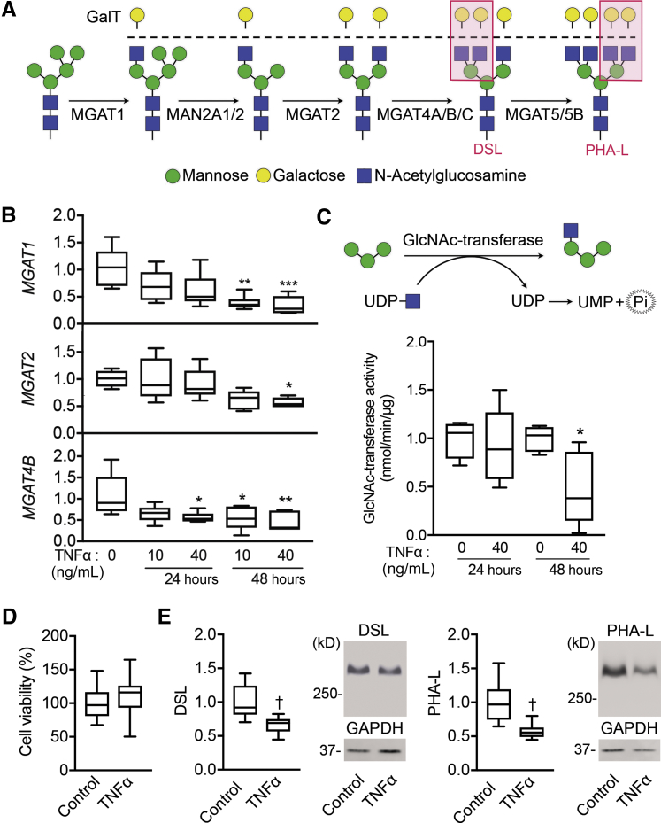
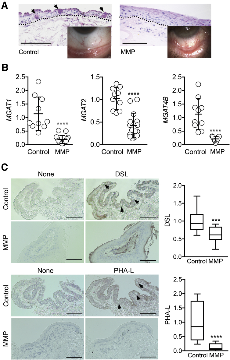
High levels of proinflammatory cytokines have been associated with a loss of tissue function in ocular autoimmune diseases, but the basis for this relationship remains poorly understood. Here we investigate a new role for tumor necrosis factor α in promoting N-glycan-processing deficiency at the surface of the eye through inhibition of N-acetylglucosaminyltransferase expression in the Golgi. Using mass spectrometry, complex-type biantennary oligosaccharides were identified as major N-glycan structures in differentiated human corneal epithelial cells. Remarkably, significant differences were detected between the efficacies of cytokines in regulating the expression of glycogenes involved in the biosynthesis of N-glycans. Tumor necrosis factor α but not IL-1β had a profound effect in suppressing the expression of enzymes involved in the Golgi branching pathway, including N-acetylglucosaminyltransferases 1 and 2, which are required for the formation of biantennary structures. This decrease in gene expression was correlated with a reduction in enzymatic activity and impaired N-glycan branching. Moreover, patients with ocular mucous membrane pemphigoid were characterized by marginal N-acetylglucosaminyltransferase expression and decreased N-glycan branching in the conjunctiva. Together, these data indicate that proinflammatory cytokines differentially influence the expression of N-glycan-processing enzymes in the Golgi and set the stage for future studies to explore the pathophysiology of ocular autoimmune diseases.


Figures

Figure 1
Figure 1
Matrix-assisted laser desorption/ionization–time-of-flight (MALDI-TOF) profile of N-glycans on glycoproteins isolated from human corneal epithelial cells. The N-linked glycans were released enzymatically by peptide:N-glycosidase F, permethylated, and profiled by MALDI-TOF mass spectrometry. Magnified portions at m/z = 1500 → 3000 (top panel), 3000 → 4100 (middle panel), and 4100 → 5200 (bottom panel) are shown. Putative structures are assigned based on compositional information and known biosynthetic pathways. Most of the N-glycans have compositions consistent with bi-antennary complex-type glycans, carrying N-acetyllactosamine residues, and high-mannose structures. All molecular ions detected are present in the form of [M + Na]+.
Figure 2
Figure 2
IL-1β and tumor necrosis factor (TNF)-α differentially affect the expression of genes involved in the processing of N-glycans. A: Relative transcript abundance of genes encoding enzymes involved in trimming and branching oligomannose, hybrid, and complex-type N-glycans in the endoplasmic reticulum and Golgi apparatus of human corneal epithelial cells. The expression of N-glycan–processing genes was assessed by real-time quantitative PCR using a human glycosylation PCR array and normalized using the 2−ΔΔCτ method with MGAT4C as calibrator. B: Scatterplot comparing the expression of N-glycan–processing genes in untreated control cells and cells treated with 10 ng/mL IL-1β or 40 ng/mL TNFα for 24 and 48 hours. The green and red dots indicate at least twofold up- or down-regulation, respectively, compared with control. Values were determined using the 2−ΔCτ method. C: Venn diagrams depicting the distribution of Golgi N-acetylglucosaminyltransferases associated with IL-1β, TNFα, or both components, at 24 and 48 hours post-treatment. Up-regulated genes are indicated in green, and down-regulated genes are indicated in red. The array data shown in these experiments were repeated twice, with independently isolated RNA pooled from three tissue culture plates. Data are expressed as means ± SD.
Figure 3
Figure 3
Tumor necrosis factor (TNF)-α reduces Golgi N-acetylglucosaminyltransferase activity and promotes loss of N-glycan branching. A: Branches in complex-type N-glycans are processed through a series of N-acetylglucosaminyltransferases and mannosidases located in the medial Golgi compartment. Red boxes indicate the GlcNAc-branched N-glycans recognized by Datura stramonium lectin (DSL) or Phaseolus vulgaris leucoagglutinin (PHA-L). B: Quantitative RT-PCR validation analysis showing reduced expression of genes involved in the formation of mono-, bi-, and tri-antennary complex-type glycans (MGAT1, MGAT2, and MGAT4B) after TNFα treatment. C:N-acetylglucosaminyltransferase activity in human corneal epithelial cells was assessed by measurement of released phosphate after the transfer of uridine diphosphate (UDP)–N-acetylglucosamine (GlcNAc) to a mannotriose acceptor substrate. D: Cell viability in stratified cell cultures treated with 40 ng/mL TNFα for 48 hours was assayed based on 3-(4,5-dimethylthiazol-2-yl)-2,5-diphenyltetrazolium bromide dye uptake (absorbance at 570 nm). E: Cell lysates from control cells and cells treated with TNFα for 72 hours were run on 1% agarose gels and then blotted to nitrocellulose membranes. Blotted proteins were probed with biotinylated lectins specific for GlcNAc-branched N-glycans (DSL and PHA-L). Representative images of the membranes are presented. Results in B–E represent at least two independent experiments performed in triplicate. The box-and-whisker plots show the 25 and 75 percentiles (boxes) and the median and the minimum and maximum data values (whiskers). Significance was determined using one-way analysis of variance with the Tukey post hoc test (B) and the t-test (C–E). P < 0.05, ∗∗P < 0.01, and ∗∗∗P < 0.001 versus 0 ng/mL TNFα; P < 0.01 versus control. GAPDH, glyceraldehyde-3-phosphate dehydrogenase; MAN, α-mannosidase; MGAT, mannoside acetylglucosaminyltransferase; UMP, uridine monophosphate.
Figure 4
Figure 4
Golgi N-acetylglucosaminyltransferase levels are substantially decreased in ocular autoimmune disease. A: Conjunctival tissue sections from control subjects and patients with ocular mucous membrane pemphigoid (MMP) were stained with periodic acid-Schiff. The arrows indicate goblet cells, whereas the dotted lines outline the basement membrane zone. The inset shows the morphologic appearance of the conjunctiva. B: Human conjunctival epithelial cells were collected in vivo via impression cytology. After total RNA extraction and reverse transcription, real-time quantitative PCR was performed using primers for MGAT1, MGAT2, and MGAT4B. C: Conjunctival tissue sections from control subjects and patients with ocular mucous membrane pemphigoid were incubated with biotin-labeled Datura stramonium lectin (DSL) or Phaseolus vulgaris leucoagglutinin (PHA-L). Consecutive sections incubated with bovine serum albumin alone were used as negative controls. The arrows indicate goblet cells. Each dot in B represents an individual biological sample, whereas quantification in C was performed by analyzing at least 10 histologic areas from three individuals in each group. The box-and-whisker plots show the 25 and 75 percentiles (boxes) and the median and the minimum and maximum data values (whiskers). Significance was determined using the t-test. Data are expressed as means ± SD. ∗∗∗P < 0.001, ∗∗∗∗P < 0.0001 versus control. Scale bars: 50 μm (A); 100 μm (C).

Similar articles

Cited by

References

    1. Apweiler R., Hermjakob H., Sharon N. On the frequency of protein glycosylation, as deduced from analysis of the SWISS-PROT database. Biochim Biophys Acta. 1999;1473:4–8. - PubMed
    1. Cummings R.D. The repertoire of glycan determinants in the human glycome. Mol Biosyst. 2009;5:1087–1104. - PubMed
    1. Moremen K.W., Tiemeyer M., Nairn A.V. Vertebrate protein glycosylation: diversity, synthesis and function. Nat Rev Mol Cell Biol. 2012;13:448–462. - PMC - PubMed
    1. Varki A. Evolutionary forces shaping the Golgi glycosylation machinery: why cell surface glycans are universal to living cells. Cold Spring Harb Perspect Biol. 2011;3:a005462. - PMC - PubMed
    1. Stanley P. Golgi glycosylation. Cold Spring Harb Perspect Biol. 2011;3:a005199. - PMC - PubMed

Publication types

MeSH terms

Substances