

Molecular mechanisms of contractile-ring constriction and membrane trafficking in cytokinesis
- PMID: 30448943
- PMCID: PMC6297088
- DOI: 10.1007/s12551-018-0479-3
Molecular mechanisms of contractile-ring constriction and membrane trafficking in cytokinesis
Abstract
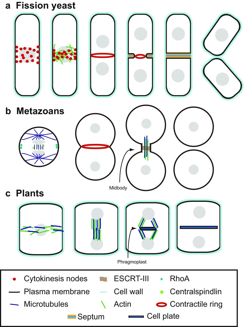
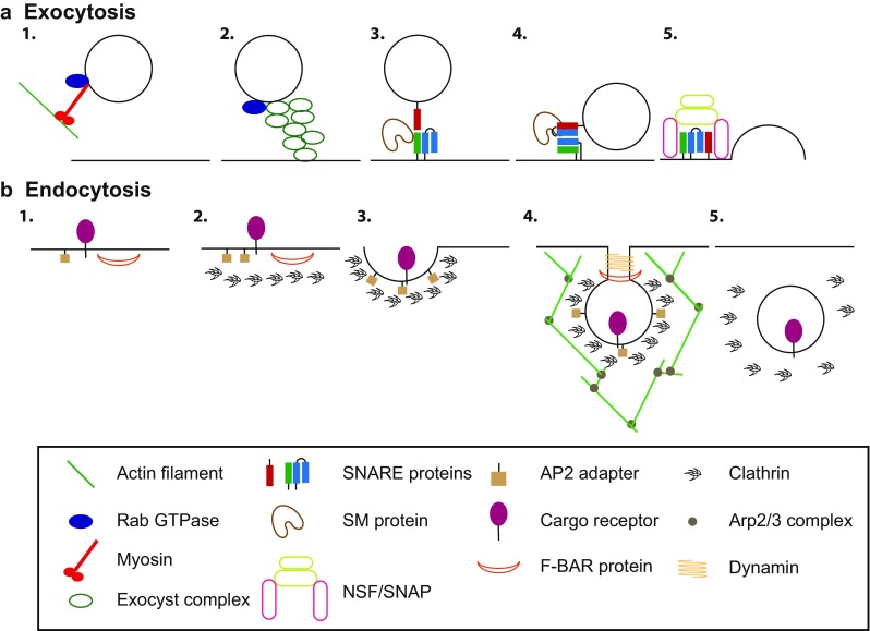
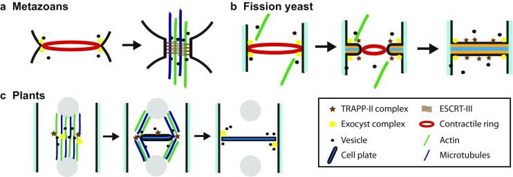
In this review, we discuss the molecular mechanisms of cytokinesis from plants to humans, with a focus on contribution of membrane trafficking to cytokinesis. Selection of the division site in fungi, metazoans, and plants is reviewed, as well as the assembly and constriction of a contractile ring in fungi and metazoans. We also provide an introduction to exocytosis and endocytosis, and discuss how they contribute to successful cytokinesis in eukaryotic cells. The conservation in the coordination of membrane deposition and cytoskeleton during cytokinesis in fungi, metazoans, and plants is highlighted.
Keywords: Contractile ring; Cytokinesis; Endocytosis; Exocytosis; Membrane deposition.
Conflict of interest statement
Conflict of interest
Kenneth S. Gerien declares that he has no conflict of interest. Jian-qiu Wu declares that he has no conflict of interest.
Ethical approval
This article does not contain any studies with human participants or animals performed by any of the authors.
Figures
Similar articles
-
Actomyosin Ring Formation and Tension Generation in Eukaryotic Cytokinesis.Curr Biol. 2016 Aug 8;26(15):R719-R737. doi: 10.1016/j.cub.2016.06.071. Curr Biol. 2016. PMID: 27505246 Review.
-
Comparing contractile apparatus-driven cytokinesis mechanisms across kingdoms.Cytoskeleton (Hoboken). 2012 Nov;69(11):942-56. doi: 10.1002/cm.21082. Epub 2012 Oct 17. Cytoskeleton (Hoboken). 2012. PMID: 23027576 Review.
-
Septation and cytokinesis in fungi.Fungal Genet Biol. 2003 Dec;40(3):187-96. doi: 10.1016/j.fgb.2003.08.005. Fungal Genet Biol. 2003. PMID: 14599886 Review.
-
[Phagocytosis and cytokinesis: highlights on common themes and differences].Med Sci (Paris). 2013 Nov;29(11):1004-9. doi: 10.1051/medsci/20132911017. Epub 2013 Nov 20. Med Sci (Paris). 2013. PMID: 24280504 Review. French.
-
The roles of the oncoprotein GOLPH3 in contractile ring assembly and membrane trafficking during cytokinesis.Biochem Soc Trans. 2015 Feb;43(1):117-21. doi: 10.1042/BST20140264. Biochem Soc Trans. 2015. PMID: 25619256
Cited by
-
Stress-activated MAPK signaling controls fission yeast actomyosin ring integrity by modulating formin For3 levels.Elife. 2020 Sep 11;9:e57951. doi: 10.7554/eLife.57951. Elife. 2020. PMID: 32915139 Free PMC article.
-
TRAPPC2l Participates in Male Germ Cell Development by Regulating Cell Division.Cell Prolif. 2025 Jun;58(6):e13810. doi: 10.1111/cpr.13810. Epub 2025 Jan 26. Cell Prolif. 2025. PMID: 40539230 Free PMC article.
-
Roles of Mso1 and the SM protein Sec1 in efficient vesicle fusion during fission yeast cytokinesis.Mol Biol Cell. 2020 Jul 15;31(15):1570-1583. doi: 10.1091/mbc.E20-01-0067. Epub 2020 May 20. Mol Biol Cell. 2020. PMID: 32432970 Free PMC article.
-
Roles of the MO25 protein Pmo25 in contractile-ring stability and localization of the NDR kinase Sid2 during cytokinesis.bioRxiv [Preprint]. 2025 May 14:2025.05.13.653815. doi: 10.1101/2025.05.13.653815. bioRxiv. 2025. PMID: 40463288 Free PMC article. Preprint.
-
Animal Cell Cytokinesis: The Rho-Dependent Actomyosin-Anilloseptin Contractile Ring as a Membrane Microdomain Gathering, Compressing, and Sorting Machine.Front Cell Dev Biol. 2020 Oct 7;8:575226. doi: 10.3389/fcell.2020.575226. eCollection 2020. Front Cell Dev Biol. 2020. PMID: 33117802 Free PMC article.
References
Publication types
Grants and funding
LinkOut - more resources
Full Text Sources
