Review
. 2019 Apr;32(4):322-330.
doi: 10.1055/s-0038-1675609. Epub 2018 Nov 16.

Diagnostic Imaging and Management of Common Intra-articular and Peri-articular Soft Tissue Tumors and Tumorlike Conditions of the Knee

Affiliations
Review

Diagnostic Imaging and Management of Common Intra-articular and Peri-articular Soft Tissue Tumors and Tumorlike Conditions of the Knee

Andrea J Evenski et al. J Knee Surg. 2019 Apr.

Abstract

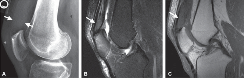
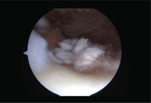
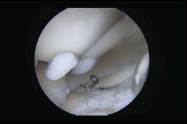
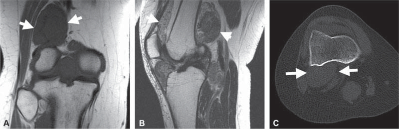
Intra-articular (IA) and peri-articular (PA) tumors of the knee are frequently encountered by orthopaedic surgeons. Nonetheless, due to the possibility of great morbidity and potential mortality, it is important to recognize and differentiate between benign and malignant lesions in a timely manner. Therefore, the purpose of this article is to provide a concise, practical, and updated review of commonly encountered IA and PA tumors including intratendinous gout, synovial chondromatosis, schwannoma, pigmented villonodular synovitis, and synovial sarcoma, and a detailed description of differentiating features to include various imaging modalities.


Conflict of interest statement

None.

Figures

Fig. 1
Fig. 1
A 50-year-old male with 3-weeks’ history of knee pain following a basketball injury referred to orthopaedic oncology for the evaluation of mass identified on magnetic resonance imaging (MRI) performed to assess for internal derangement. (A) Lateral radiographs demonstrate thickening of the quadriceps tendon (white arrows), erosion of a superior patellar enthesophyte (arrowhead), and faint mineralization in Hoffa’s fat (asterisks). (B) Sagittal T2-weighted image with fat suppression and (C) T1-weighted image demonstrates a mass infiltrating the quadriceps tendon (white arrows in B and C) with intermediate T2 and hypointense T1 signal and reactive marrow edema in the patella compatible with the gout. The diagnosis was confirmed on subsequent ultrasound-guided needle biopsy.
Fig. 2
Fig. 2
Arthroscopic image displaying pedunculated nature of synovial chondromatosis seen in phase I and II.
Fig. 3
Fig. 3
Arthroscopic image showing loose osteochondral bodies representative of synovial chondromatosis.
Fig. 4
Fig. 4
A 30-year-old female presenting with a 3-year history of progressive knee pain. (A) Coronal T1-weighted and (B) sagittal T2-weighted images without fat suppression demonstrate a heterogeneous synovial based mass (white arrows in A-C) distending thejoint space without bone erosion. There were no foci of signal void or intralesional fat, as demonstrated onT1-weighted image. No calcified or ossified mass was seen on the subsequent axial computed tomography (CT) in bone window (C), indicative of primary synovial chondromatosis pattern A.
Fig. 5
Fig. 5
Biopsy specimen demonstrating Antoni A and B areas consistent with nerve sheath tumor.
Fig. 6
Fig. 6
Diffuse uptake for S100 consistent with schwannoma.
Fig. 7
Fig. 7
A 50-year-old man underwent knee magnetic resonance imaging (MRI) for anterior knee pain and was referred for the evaluation of incidental mass in the popliteal fossa. (A) Axial T2-weighted image with fat suppression shows an encapsulated T2 hyperintense mass (white arrow) with smooth margins posterior to the popliteal artery (asterisks) inseparable from the tibial nerve and compatible with a schwannoma. (B) Coronal T1-weighted image shows the hypointense mass with the fascicular appearance of the tibial nerve at the proximal and distal aspects of the mass. (C) Sagittal T2-weighted image shows the “target sign,” (white arrow) low central signal intensity and high signal intensity in the periphery, an indicator of a benign peripheral nerve sheath tumor.
Fig. 8
Fig. 8
A 34-year-old with knee mass identified on magnetic resonance imaging (MRI) performed for pain following a twisting injury. (A) Lateral knee radiograph shows a soft tissue attenuation mass (black arrows) in Hoffa’s fat without calcification. (B) Axial T2-weighted image with fat suppression and (C) coronal T1-weighted image through the anterior knee show a mass (white arrows in B and C) that has heterogeneous T2 signal hypointensity and homogeneous T1 signal hypointensity. (D) Sagittal gradient echo image shows “blooming artifact” (white arrowhead) at the periphery of the mass, diagnostic of focal nodular synovitis. The “blooming” at the margins is due to local changes in the magnetic field induced by the ferromagnetic effects of hemosiderin deposited in the tumor.
Fig. 9
Fig. 9
A 58-year-old man presented with enlarging anterior knee mass following impact injury 1 to 2 years prior. Mass was previously stable for many years and initially mistaken for a cyst. (A) Axial T2-weighted image with fat suppression demonstrates a large juxta-articular mass (white arrows in A-C) with triple signal intensity, areas of high, and intermediate and low signal intensity. (B) On coronal T1-weighted image, the mass is sharply marginated with smooth round contours and heterogeneous signal. (C) The mass enhances heterogeneously post contrast on sagittal T1-weighted image with fat suppression excluding cyst from the differential diagnosis. The diagnosis of synovial sarcoma was confirmed on computed tomography guided biopsy.

Similar articles

Cited by

References

    1. Cronin KA, Lake AJ, Scott S, et al. Annual Report to the Nation on the Status of Cancer, part I: National cancer statistics. Cancer 2018;124(13):2785–2800 - PMC - PubMed
    1. Smith E, Hoy DG, Cross M, et al. The global burden of other musculoskeletal disorders: estimates from the Global Burden of Disease 2010 study. Ann Rheum Dis 2014;73(08):1462–1469 - PubMed
    1. Rodas G, Pedret C, Catala J, Soler R, Orozco L, Cusi M. Intratendi- nous gouty tophus mimics patellar tendonitis in an athlete. J Clin Ultrasound 2013;41(03):178–182 - PubMed
    1. Colberg RE, Henderson RG. Diagnosis and treatment of gouty tophi in the patellar tendon using ultrasound-guided needle barbotage: a case presentation. PM R 2017;9(09):938–942 - PubMed
    1. Gililland JM, Webber NP, Jones KB, Randall RL, Aoki SK. Intraten- dinous tophaceous gout imitating patellar tendonitis in an athletic man. Orthopedics 2011;34(03):223 - PMC - PubMed

MeSH terms