. 2018 Dec 20;72(6):999-1012.e6.
doi: 10.1016/j.molcel.2018.10.012. Epub 2018 Nov 15.

Cryo-EM Structures of MDA5-dsRNA Filaments at Different Stages of ATP Hydrolysis

Affiliations

Cryo-EM Structures of MDA5-dsRNA Filaments at Different Stages of ATP Hydrolysis

Qin Yu et al. Mol Cell. .

Abstract

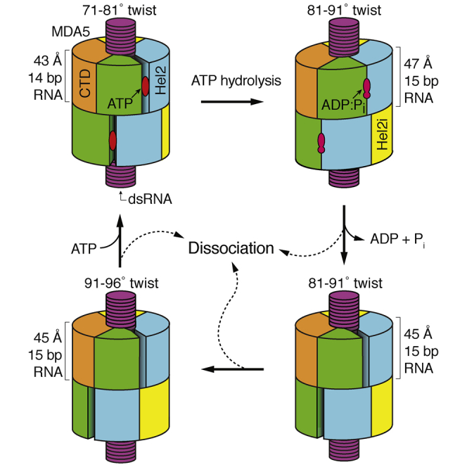
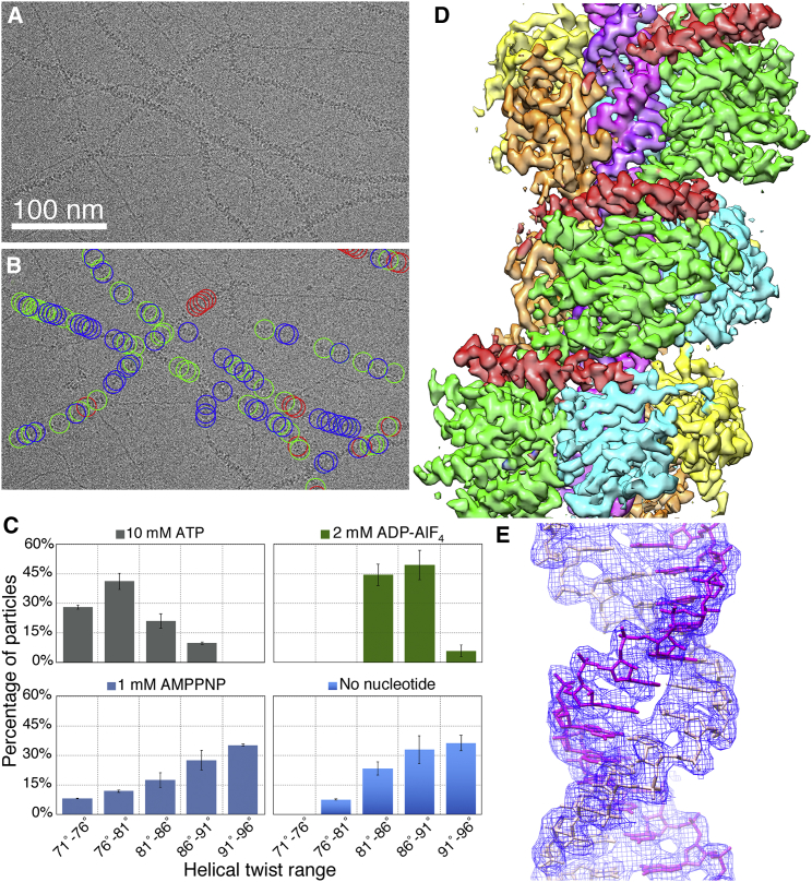
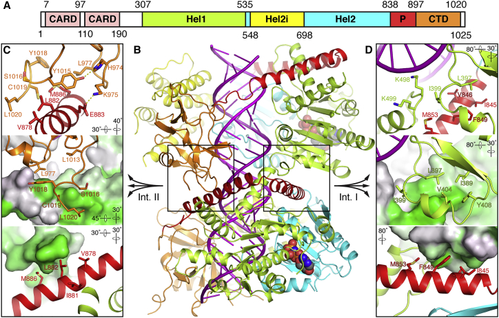
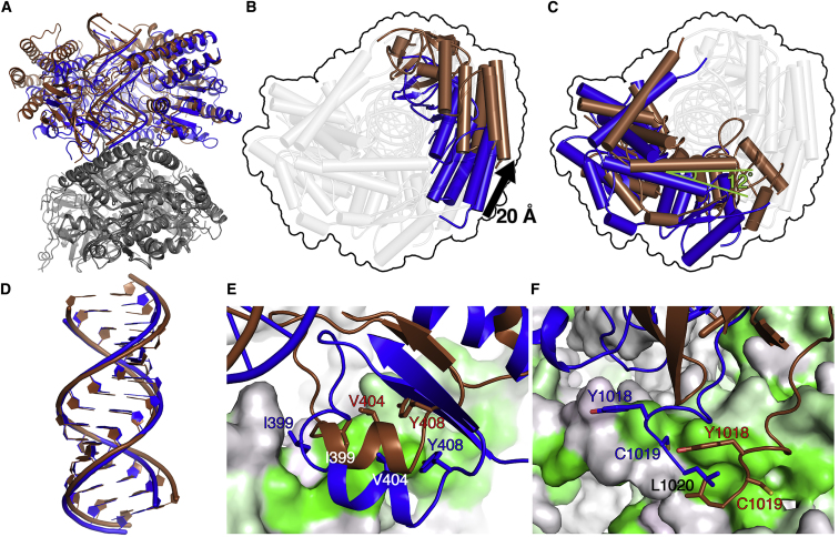
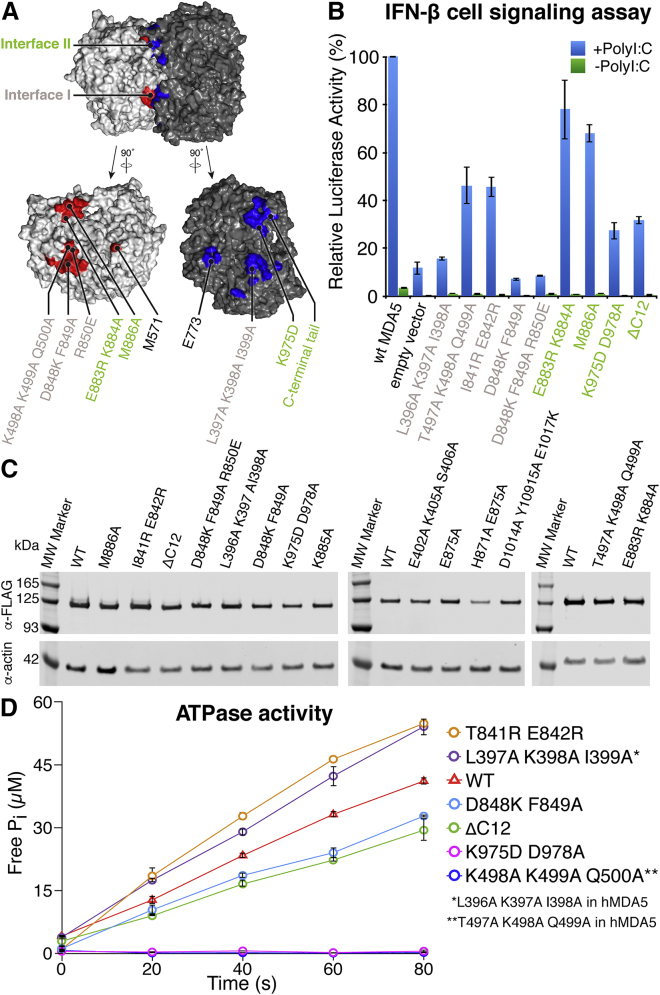
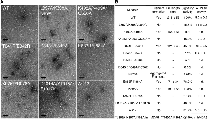
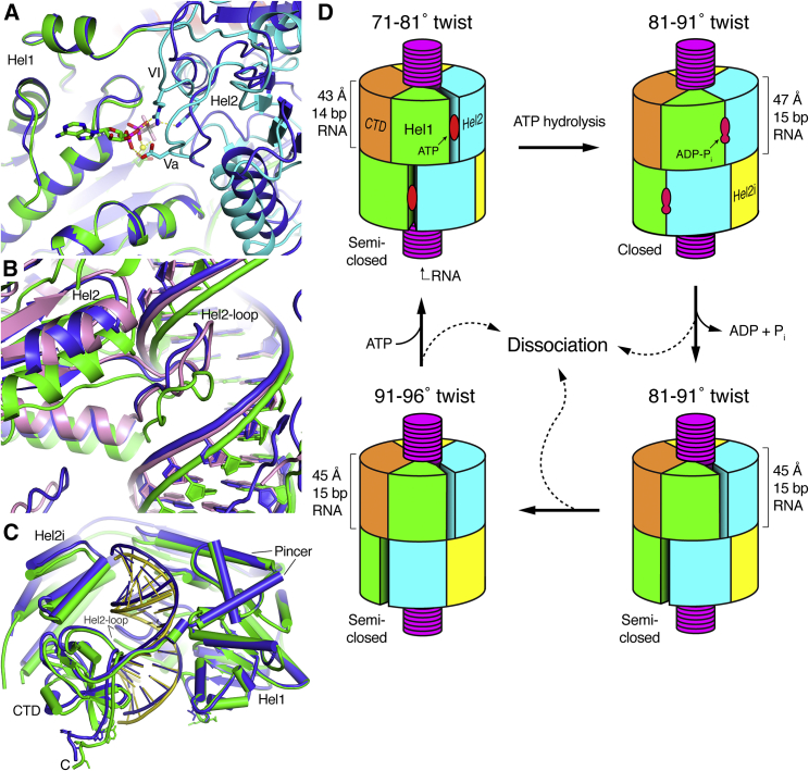
Double-stranded RNA (dsRNA) is a potent proinflammatory signature of viral infection. Long cytosolic dsRNA is recognized by MDA5. The cooperative assembly of MDA5 into helical filaments on dsRNA nucleates the assembly of a multiprotein type I interferon signaling platform. Here, we determined cryoelectron microscopy (cryo-EM) structures of MDA5-dsRNA filaments with different helical twists and bound nucleotide analogs at resolutions sufficient to build and refine atomic models. The structures identify the filament-forming interfaces, which encode the dsRNA binding cooperativity and length specificity of MDA5. The predominantly hydrophobic interface contacts confer flexibility, reflected in the variable helical twist within filaments. Mutation of filament-forming residues can result in loss or gain of signaling activity. Each MDA5 molecule spans 14 or 15 RNA base pairs, depending on the twist. Variations in twist also correlate with variations in the occupancy and type of nucleotide in the active site, providing insights on how ATP hydrolysis contributes to MDA5-dsRNA recognition.

Keywords: AGS; ATPase; Aicardi-Goutières syndrome; DExD/H-box RNA helicase; RIG-I-like receptor; RLR; SF2 helicases; SMS; Singleton-Merten syndrome; cryo-EM; cryoelectron microscopy; helical reconstruction; innate immune pattern recognition; nucleic acid sensing; superfamily 2 helicases.


Figures

None
Graphical abstract
Figure 1
Figure 1
Cryo-EM Image Reconstruction of MDA5-dsRNA Filaments with Helical Symmetry Averaging (A) Representative cryo-EM micrograph of MDA5-dsRNA filaments. (B) Cryo-EM micrograph shown in (A) with circles drawn around the boxed filament segments that were used in the helical reconstructions. The circles are colored according to the 3D class that they contributed to. Segments that contributed to the Twist74, Twist87, and Twist91 structures are in red, green, and blue, respectively. (C) Histogram showing the distributions of filament segments as a function of helical twist for the ATP, ADP-AlF4, 1-mM AMPPNP, and nucleotide-free datasets. The distributions shown are from 3D classification performed with ten classes per dataset. Error bars represent SEM between replicate 3D classification calculations; n = 3. (D) 3D density map of the Twist74 MDA5-dsRNA filament at 3.68 Å overall resolution. The components are colored as follows: Hel1, green; Hel2, cyan; Hel2i, yellow; pincer domain, red; CTD, orange; and RNA, magenta. (E) The dsRNA density in the Twist74 filament (blue mesh) is shown with the fitted atomic model (magenta and pink). See also Figures S1 and S2.
Figure 2
Figure 2
Atomic Model of the MDA5-dsRNA Filament (A) Domain structure of mouse MDA5. CARD, caspase recruitment domain; CTD, C-terminal domain; Hel1 and Hel2, first and second RecA-like helicase domains; Hel2i, Hel2 insert domain; P, pincer domain. The same color code and domain abbreviations are used in subsequent panels and in Figures 1, 7A, and 7D. (B) Overview of the refined atomic model of the MDA5-dsRNA filament. Two adjacent MDA5 subunits and 28 bp of dsRNA are shown from the Twist74 structure. RNA is in magenta. The bound AMPPNP molecules are shown in sphere representation. The two filament-forming interfaces are boxed. (C and D) Close-up views of filament interface II (C) and interface I (D). The top panels show side chains forming key contacts, with hydrogen bonds shown as yellow dashed lines. In the middle panels the lower protomer in (B) is shown in surface representation colored by hydrophobicity from gray to green, with green being the most hydrophobic. In the lower panels, the upper protomer in (B) is shown in surface representation colored by hydrophobicity. The orientation of the view relative to (B) is indicated for each panel. See also Figure S3 and Videos S1, S2, S3, S4, and S5.
Figure 3
Figure 3
Close-up Views of the Cryo-EM Densities and Atomic Models around the ATP-Binding Site for Reconstructions with Different Helical Twists and Bound Nucleotides (A–C) Density consistent with a nucleotide triphosphate molecule is visible in the low-twist structures with 2.5 mM AMPPNP (B) and 10 mM ATP (C), but only weak density is visible in the low-twist (74°) 1 mM AMPPNP structure (A) (red outline). (D) With 2 mM ADP-AlF4, strong density is visible for the ADP and AlF4 moieties (green box). The AlF4 moiety shown in pink and gray and a coordinated Mg2+ ion in cyan. (E–H) With 1–2.5 mM AMPPNP (E and F) or no nucleotide (G and H), there is no nucleotide density in the catalytic site of the structures with mid- to high helical twist (81°–96°, blue box). A contour level of 4.5 σ in PyMol was used for all panels. The AMPPNP, ATP, and ADP-AlF4 molecules and selected protein side chains are shown in stick representation. See also Figure S3.
Figure 4
Figure 4
Differences in Relative Domain Positions and Filament Contacts in the Twist74 and Twist91 Structures (A) Overview of two protomers of the Twist74 and Twist91 structures superimposed using the pincer domain of the lower protomer (gray) as the reference. The upper protomer of Twist74 is in blue and that of Twist91 is in brown. (B and C) Top views along the helical axis of the upper protomer from (A) showing the shifts in the positions of Hel2i and CTD (B), and Hel1, Hel2, and pincer (C). The 20 Å translation in Hel2i and 12° rotation in the pincer domain are highlighted. The highlighted domains are colored as in the upper protomer in (A), and the remaining domains are shown in transparent gray for clarity. The outer contour of the superimposed structures is shown for reference as a black outline. (D) Close-up of the dsRNAs from the Twist74 and Twist91 structures from the structural alignment in (A). The RMSD of the atoms in the 14 superimposed RNA base pairs is 1.23 Å. (E and F) Close-up views of filament interface I (E) and interface II (F). The protomer of Twist74 shown in gray in (A) and used as the alignment reference is shown in surface representation colored by hydrophobicity as in Figure 2. Key interface residues in the adjacent protomer are shown with Twist74 in blue and Twist91 in brown. See also Figures S4–S6 and Videos S1, S2, S3, S4, and S5.
Figure 5
Figure 5
Mutations at the Filament-Forming Interfaces Abolish or Reduce Cell Signaling in Response to dsRNA (A) Location of the engineered filament interface mutations. Two filament protomers are shown in surface representation with the filament-forming surfaces of each protomer colored in red and blue, respectively. The protomers are shown assembled with the helical axis horizontal (top) and separately after being opened like a book with 90° rotations in opposite directions to show the interface surfaces (bottom). Residue labels are colored pink for interface I and green for interface II. Residue numbers refer to mouse MDA5. (B) IFN-β reporter cell signaling assay. Plasmids encoding human MDA5 mutants were co-transfected into HEK293 cells with plasmids encoding firefly luciferase under an IFN-β-inducible promoter and Renilla luciferase under a constitutive promoter. Cells were later transfected with poly(I:C) RNA (+PolyI:C) or DMEM (−PolyI:C). Relative luciferase activity was calculated as the ratio of firefly to Renilla luciferase luminescence. Residue numbers refer to human MDA5. Error bars represent SEM between measurements; n = 3. (C) Western blots showing the expression level of the human MDA5 mutants in HEK293T cells. The FLAG tag on each MDA5 variant was detected with an anti-FLAG antibody. (D) ATP hydrolysis assay. The ATPase activities of MDA5 mutants with reduced signaling activity were measured as release of inorganic phosphate (Pi) on incubation with ATP and 1-kb dsRNA. Error bars represent SEM; n = 3. See also Figure S7.
Figure 6
Figure 6
Interface Mutations that Impair Signaling Also Impair Filament Formation (A) Representative electron micrographs of MDA5 filament interface mutants in the presence of 1 kb dsRNA, 1 mM AMPPNP, and 5 mM MgCl2. Scale bars, 100 nm. Residue numbers refer to mouse MDA5. (B) Table summarizing the filament formation activity, filament length, cell-signaling activity, and ATPase activity of selected MDA5 mutants. ATPase activities were calculated from the initial slopes of the curves in Figure 5D and is expressed as moles of released phosphate per mole of MDA5 per second (MPi MMDA5−1 s−1). Residue numbers refer to mouse MDA5. For mutants with different residue numbers in human MDA5, the corresponding mutation is shown in human residue numbers at the bottom. n.d., not determined. See also Figures S5 and S7.
Figure 7
Figure 7
Comparison of the Closed ADP-AlF4-Bound Structure with the Semi-open Structures and Schematic Model of the ATPase Cycle and Proofreading Mechanism of MDA5 For a Figure360 author presentation of Figure 7, see https://doi.org/10.1016/j.molcel.2018.10.012. (A) Close-up view of the nucleotide-binding site and Hel1-Hel2 domain interface. The Twist74 AMPPNP-bound structure (blue) was superimposed on the ADP-AlF4-bound structure (colored by domain as in Figure 2) using the Hel1 domain as the reference. Nucleotide-binding motifs Va and VI are labeled. Only the ADP-AlF4 nucleotide is shown for clarity. (B) Close-up view of the Hel2-loop and its interactions with the dsRNA. The Twist74 (blue) and Twist87 (pink) AMPPNP-bound structures are superimposed onto the ADP-AlF4-bound structure (green) using Hel1 as the reference. (C) Overview of Twist74 (blue) superimposed on the ADP-AlF4-bound structure (green) using Hel1 as the reference. (D) Model of the ATPase cycle and proofreading mechanism. Only two filament protomers are shown for clarity. The low-twist (71°–81°) structures correspond to the ATP-bound catalytic ground state, the intermediate-twist (81°–91°) ADP-AlF4-bound structure is the transition state, and the intermediate- and high-twist (91°–96°) states represent nucleotide-free states. The four panels relate to the panels in Figures 3C–3F. See also Videos S1, S2, S3, S4, and S5.

References

    1. Adams P.D., Afonine P.V., Bunkóczi G., Chen V.B., Davis I.W., Echols N., Headd J.J., Hung L.W., Kapral G.J., Grosse-Kunstleve R.W. PHENIX: a comprehensive Python-based system for macromolecular structure solution. Acta Crystallogr. D Biol. Crystallogr. 2010;66:213–221. - PMC - PubMed
    1. Ahmad S., Mu X., Yang F., Greenwald E., Park J.W., Jacob E., Zhang C.Z., Hur S. Breaching self-tolerance to Alu duplex RNA underlies MDA5-mediated inflammation. Cell. 2018;172:797–810.e713. - PMC - PubMed
    1. Amunts A., Brown A., Bai X.C., Llácer J.L., Hussain T., Emsley P., Long F., Murshudov G., Scheres S.H.W., Ramakrishnan V. Structure of the yeast mitochondrial large ribosomal subunit. Science. 2014;343:1485–1489. - PMC - PubMed
    1. Aricescu A.R., Lu W., Jones E.Y. A time- and cost-efficient system for high-level protein production in mammalian cells. Acta Crystallogr. D Biol. Crystallogr. 2006;62:1243–1250. - PubMed
    1. Berke I.C., Modis Y. MDA5 cooperatively forms dimers and ATP-sensitive filaments upon binding double-stranded RNA. EMBO J. 2012;31:1714–1726. - PMC - PubMed

Publication types

MeSH terms