. 2018 Dec 12;24(6):804-816.e6.
doi: 10.1016/j.chom.2018.10.015. Epub 2018 Nov 15.

Essential cGMP Signaling in Toxoplasma Is Initiated by a Hybrid P-Type ATPase-Guanylate Cyclase

Affiliations

Essential cGMP Signaling in Toxoplasma Is Initiated by a Hybrid P-Type ATPase-Guanylate Cyclase

Kevin M Brown et al. Cell Host Microbe. .

Abstract

Apicomplexan parasites rely on cyclic nucleotide-dependent kinases for host cell infection, yet the mechanisms that control their activation remain unknown. Here we show that an apically localized guanylate cyclase (GC) controls microneme secretion and lytic growth in the model apicomplexan Toxoplasma gondii. Cell-permeable cGMP reversed the block in microneme secretion seen in a knockdown of TgGC, linking its function to production of cGMP. TgGC possesses an N-terminal P-type ATPase domain fused to a C-terminal heterodimeric guanylate cyclase domain, an architecture found only in Apicomplexa and related protists. Complementation with a panel of mutants revealed a critical requirement for the P-type ATPase domain for maximum GC function. We further demonstrate that knockdown of TgGC in vivo protects mice from lethal infection by blocking parasite expansion and dissemination. Collectively, this work demonstrates that cGMP-mediated signaling in Toxoplasma relies on a multi-domain architecture, which may serve a conserved role in related parasites.

Keywords: apicomplexan parasite; auxin-inducible degron; calcium; cyclic nucleotides; egress; host-pathogen interaction; invasion; regulated protein stability; secretion; signaling.


Figures

Figure 1-
Figure 1-
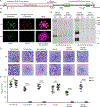
Reverse genetic screen for essential purine nucleotide cyclases in T. gondii (A) Predicted protein domain architectures of the five putative purine nucleotide cyclases in T. gondii based on NCBI CDD search.(B) Alignment of purine binding pockets of selected eukaryotic nucleotide cyclases. Residues important for purine selectivity (adenine = blue, guanine = yellow) and catalysis (green) are highlighted. The original alignment (Baker and Kelly, 2004) was modified here to include HuADCY2, PfGCα, and Toxoplasma cyclases. See Table S1 for protein accessions used in the alignment.(C) Plaques formed by wild type and knockout mutants on HFF monolayers at D8, 100 parasites / monolayer. Data represented as mean ± SD (n = 9-12 replicates combined from N = 3 or 4 trials). One-way ANOVA with Dunnett’s multiple comparisons test vs parent, Adjusted P values: * ≤ 0.05; *** ≤ 0.001; **** ≤ 0.0001. See also Figures S1A, S1B.(D) Schematic representation of taxa with gene or transcript evidence of a P-type ATPase/guanylate cyclase. See also Figure S1C and Table S1.
Figure 2-
Figure 2-
TgGC is essential for the lytic life cycle of T. gondii (A) Domain architecture of TgGC showing functional (NCBI CDD) and transmembrane domains (OCTOPUS) as well as insertion points for N- and C-terminal tags. See also Figure S2A. (B) IF microscopy of intracellular RH 6Ty-GC-mAID-3HA parasites treated with indole acetic acid (IAA) or vehicle for 14 h. Parasites were labeled with mouse anti-Ty and anti-mouse IgG Alexa Fluor 488 (green), rat anti-HA and anti-rat IgG Alexa Fluor 568 (red), and rabbit anti-TgGAP45 and anti-rabbit IgG Alexa Fluor 647 (magenta). Scale bar = 5 μm. See also Figure S2B. (C) Immunoblot of lysates from RH TIR1-3FLAG and derivative RH 6Ty-GC-mAID-3HA parasites treated with IAA or vehicle for 14 h. Blots were probed with rabbit anti-Ty and anti-rabbit IgG IRDye 680RD and mouse anti-HA and anti-mouse IgG IRDye 800CW. Separate channels of the same membrane scan are shown. See also Figure S2C. (D-E) Plaques formed by TIR1-3FLAG and derivative TgGC tagged lines on HFF monolayers with IAA or vehicle control (Ctrl) at D8 with 200 parasites/ monolayer. Scale bar = 5 mm. (E) Plaque data represented as mean ± SD (n = 9 replicates combined from N = 3 trials). Each parasite line was analyzed individually for statistical significance using an unpaired t test (IAA vs control), P values: **** ≤ 0.0001.
Figure 3-
Figure 3-
TgGC regulates PKG-dependent microneme secretion for motile processes (A-B) Replication of RH TIR1-3FLAG and derivative RH GC-mAID-3HA parasites grown in HFFs for 24 h in the presence of IAA or vehicle (added 1 h post-invasion). See also Figure S3A. (A) Mean percentages of parasite vacuoles containing indicated numbers of parasites ± SEM (N = 3). (B) Mean parasites per vacuole ± SEM (N = 3). Each parasite line was analyzed individually for statistical significance using an unpaired t test (IAA vs control). ns = not significant. (C) Egress assay of RH TIR1-3FLAG and RH GC-mAID-3HA parasites grown for 40 h in HFFs in the presence of IAA or vehicle (administered at 26 h post-infection). At 40 h post-infection, cultures were given a 5 min pulse with A23187 or vehicle. Shown are the percentages of intact and egressed vacuoles based on IF microscopy from a one of three trials (N = 3) with similar outcomes. See also Figure S3B. (D) Invasion of RH TIR1-3FLAG and RH GC-mAID-3HA parasites on HFF monolayers following 14 h treatment with IAA or vehicle. Data represented as mean ± SEM (N = 3). Each parasite line was analyzed individually for statistical significance using an unpaired t test (IAA vs control), P values: **** ≤ 0.0001. See also Figure (E) Motility of RH TIR1-3FLAG and RH GC-mAID-3HA parasites on glass coverslips following 14 h treatment with IAA or vehicle. Motility trails were detected by IF microscopy using rabbit anti-TgSAG1 and anti-rabbit IgG Alexa Fluor 488. Representative fields from one of three experiments with similar outcome are shown. (F-G) Microneme secretion of extracellular RH TIR1-3FLAG and RH GC-mAID-3HA parasites stimulated ± serum albumin/ethanol following 14 h treatment with IAA or vehicle. Stim = stimulated, mock = mock stimulation with vehicle. (F) Western blot of T. gondii excreted/secreted antigens probed for secreted micronemes (anti-MIC2) and constitutive dense granule secretion (anti-GRA7). Separate channels of each membrane scan (Stim, Mock) are shown. (G) Quantification of MIC2 secretion detected by Western blotting using densitometry. Data represented as mean ± SD (N = 3). Each parasite line and stimulation condition were analyzed individually for statistical significance using an unpaired t test (IAA vs control), P values: * < 0.01. (H) Microneme secretion of extracellular RH TIR1-3FLAG, RH GC-mAID-3HA, and RH PKG-mAID-3HA parasites stimulated ± PET-cGMP or PET-cGMP + Zaprinast following 14 h treatment with IAA. Western blot of T. gondii excreted/secreted antigens probed for secreted micronemes (anti-MIC2) and constitutive dense granule secretion (anti-GRA7). Separate channels of the same membrane scan are shown. Representative of four experiments with the same outcome. (I) Model of TgGC-dependent microneme secretion.
Figure 4-
Figure 4-
Expression and localization of mutant TgGC complementation constructs (A) Domain architectures of wild-type and mutant TgGC-mCherry fusion constructs. (B) IF microscopy of extracellular RH GC-mAID-3HA parasites stably expressing second copies of TgGC-mCherry fusions (depicted in Figure 4A, matched rows) probed for TgGC fusions (rat anti-mCherry and anti-rat IgG Alexa Fluor 568) and the apical cap (mouse anti-TgISP1 and anti-mouse IgG Alexa Fluor 488). Scale bar = 5 μm. Identical immunolabeling and imaging protocols were used for each line. The mean fluorescent intensities (MFI) of the raw mCherry signal data were averaged from at least 30 parasites (mean ± SD) for each line from 1 of 3 independent experiments with similar outcomes. Statistical differences in MFIs were assessed by one-way ANOVA with Dunnett’s multiple comparisons test (mutant vs wild-type control), adjusted P values are shown. See also Figures S4A-D.
Figure 5-
Figure 5-
Functional analysis of the multi-domain structure of TgGC (A) Microneme secretion of extracellular RH GC-mAID-3HA complement lines. Parasites were stimulated ± serum albumin/ethanol following 14 h treatment with IAA or vehicle. Western blot of T. gondii excreted/secreted antigens probed for secreted micronemes (anti-MIC2) and constitutive dense granule secretion (anti-GRA7). Stim = stimulated, mock = mock stimulation with vehicle. Representative blot shown (N = 2 similar experiments). (B) Plaques formed by RH GC-mAID-3HA complement lines on HFF monolayers with IAA or vehicle at D8, 200 parasites/ monolayer. Plaque data represented as mean ± SD (n = 9 replicates combined from N = 3 trials). Each parasite line was analyzed individually for statistical significance using an unpaired t test (IAA vs control), P values: **** ≤ 0.0001.
Figure 6-
Figure 6-
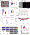
Depletion of TgGC in vivo protects mice from lethal toxoplasmosis (A) IF microscopy of Me49 TIR1-3FLAG and Me49 GC-mAID-3HA treated with IAA or vehicle for 14 h. Parasites were labeled with rat anti-FLAG and anti-rat IgG Alexa Fluor 568 and mouse anti-HA and anti-mouse IgG Alexa Fluor 488. Scale bar = 5 μm. See also Figures S5A-D. (B) Plaques formed by Me49 FLuc, Me49 TIR1-3FLAG, and Me49 GC-mAID-3HA parasite lines on HFF monolayers with IAA or vehicle at D8, 200 parasites/ monolayer. Plaque data represented as mean ± SD (n = 15 replicates combined from N = 3 trials). Each parasite line was analyzed individually for statistical significance using an unpaired t test (IAA vs control), P values: **** ≤ 0.0001. (C) In vivo assessment of TgGC-mAID-3HA knockdown. C57Bl/6 mice were challenged with 1000 Me49 GC-mAID-3HA parasites intraperitoneally and treated with IAA or vehicle on days 5-6. On day 6, mice were sacrificed, and peritoneal exudate cells were collected for IF microscopy. Fixed cells were probed for parasites (rabbit anti-TgGAP45 and anti-rabbit IgG Alexa Fluor 647) and GC-mAID-3HA (mouse anti-HA and antimouse IgG Alexa Fluor 488). Scale bar = 5 μm. Identical immunolabeling and imaging protocols were used for each treatment group. (D) Quantification TgGC-mAID-3HA and TgGAP45 expression depicted in (C). The abundance of each protein (mean fluorescence intensity ± SD) was quantified and averaged from n = 20 parasites for each treatment group from one of two trials (N = 2). Statistical significance was assessed using an unpaired t test (IAA vs control), P values: **** ≤ 0.0001. n.s. = not significant. (E) Experimental design of in vivo test of TgGC essentiality using AID. Related to Figures 6F-J (n = 9 to 10 mice per group combined from the same N = 2 trials). (F) Survival curve of C57Bl/6 mice infected with 200 Me49 GC-mAID-3HA parasites intraperitoneally and treated with IAA or vehicle for 15 days. The Gehan-Breslow- Wilcoxon test was used to compare differences between the survival curves, P < 0.0001 (IAA vs Control). (G) Mean body weight ± SD of C57Bl/6 mice infected with 200 Me49 GC-mAID-3HA parasites intraperitoneally and treated with IAA or vehicle for 15 days. (H-I) Bioluminescence imaging of C57Bl/6 mice infected i.p. with 200 Me49 GC-mAID- 3HA parasites then treated with IAA or vehicle for 15 days. (H) Radiance overlay of luciferase-expressing Me49 GC-mAID-3HA in C57BI/6 mice at days 6, 9, 12, and 37 post-infection. (I) Quantification of whole body radiance shown in (H). Line indicates mean radiance. (J) Tissue cysts per brain from C57BI/6 mice infected with 200 Me49 GC-mAID-3HA parasites intraperitoneally and treated with IAA 15 days, then euthanized on day 40 post-infection for cyst determination in brain homogenates by microscopy. See also Figures S5E, S5F.

Similar articles

Cited by

References

    1. Alaganan A, Fentress SJ, Tang K, Wang Q, and Sibley LD (2013). Toxoplasma GRA7 effector increases turnover of immunity-related GTPases and contributes to acute virulence in the mouse. Proc Natl Acad Sci (USA) 111, 1126–1131. - PMC - PubMed
    1. Baker DA, Drought LG, Flueck C, Nofal SD, Patel A, Penzo M, and Walker EM (2017). Cyclic nucleotide signalling in malaria parasites. Open Biol 7, 10.1098/rsob.170213. - DOI - PMC - PubMed
    1. Baker DA, and Kelly JM (2004). Structure, function and evolution of microbial adenylyl and guanylyl cyclases. Mol Microbiol 52, 1229–42. - PubMed
    1. Bastin P, Bagherzadeh Z, Matthews KR, and Gull K (1996). A novel epitope tag system to study protein targeting and organelle biogenesis in Trypanosoma brucei. Molec Biochem Parasitol 77, 235–239. - PubMed
    1. Beck JR, Rodriguez-Fernandez IA, de Leon JC, Huynh MH, Carruthers VB, Morrissette NS, and Bradley PJ (2010). A novel family of Toxoplasma IMC proteins displays a hierarchical organization and functions in coordinating parasite division. PLoS Pathog 6, e1001094. - PMC - PubMed

Publication types