. 2018 Nov 18;10(11):3327-3352.
doi: 10.18632/aging.101643.

Oxidative muscles have better mitochondrial homeostasis than glycolytic muscles throughout life and maintain mitochondrial function during aging

Affiliations

Oxidative muscles have better mitochondrial homeostasis than glycolytic muscles throughout life and maintain mitochondrial function during aging

Annunziata N Crupi et al. Aging (Albany NY). .

Abstract

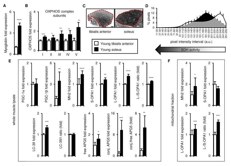
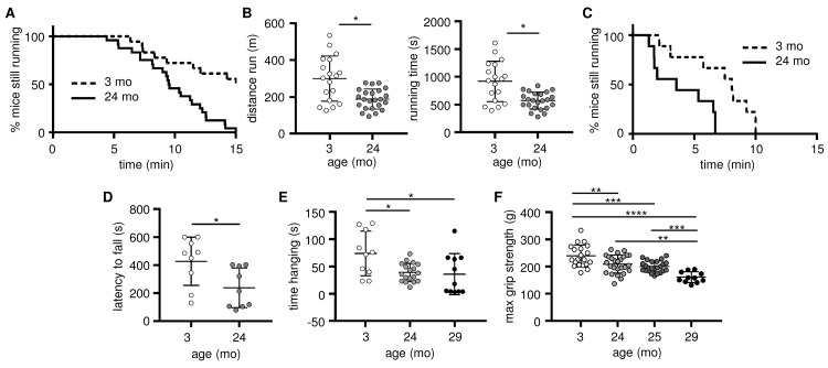
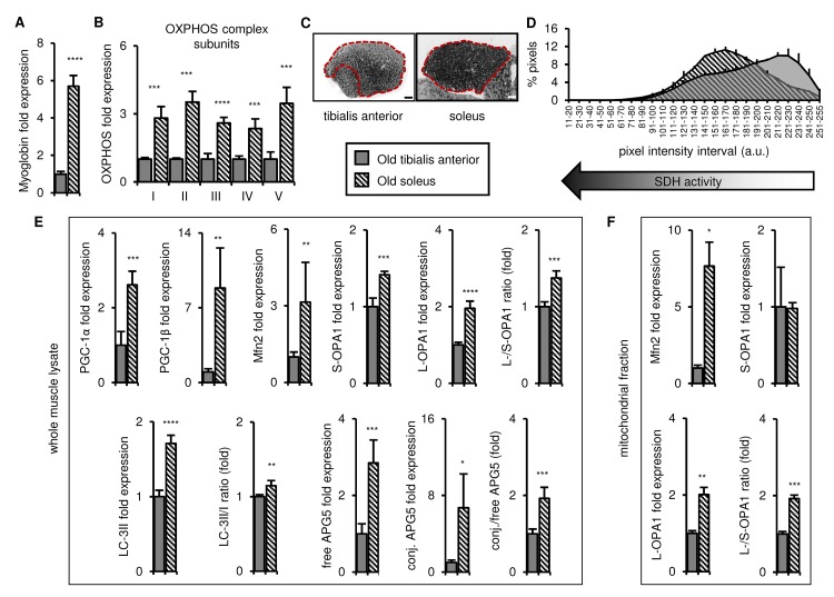
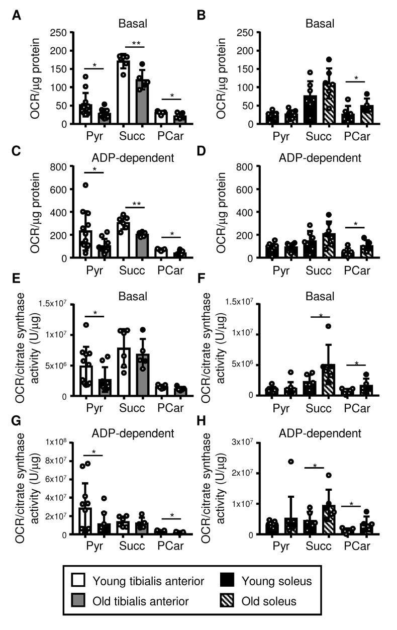
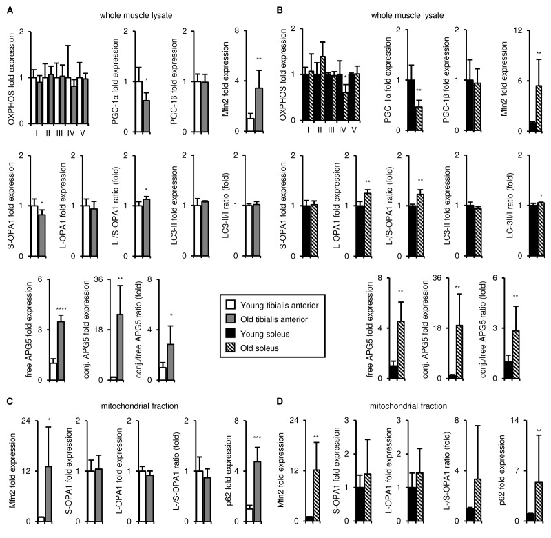
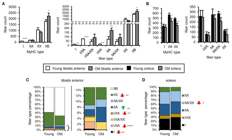
Preservation of mitochondrial function, which is dependent on mitochondrial homeostasis (biogenesis, dynamics, disposal/recycling), is critical for maintenance of skeletal muscle function. Skeletal muscle performance declines upon aging (sarcopenia) and is accompanied by decreased mitochondrial function in fast-glycolytic muscles. Oxidative metabolism promotes mitochondrial homeostasis, so we investigated whether mitochondrial function is preserved in oxidative muscles. We compared tibialis anterior (predominantly glycolytic) and soleus (oxidative) muscles from young (3 mo) and old (28-29 mo) C57BL/6J mice. Throughout life, the soleus remained more oxidative than the tibialis anterior and expressed higher levels of markers of mitochondrial biogenesis, fission/fusion and autophagy. The respiratory capacity of mitochondria isolated from the tibialis anterior, but not the soleus, declined upon aging. The soleus and tibialis anterior exhibited similar aging-associated changes in mitochondrial biogenesis, fission/fusion, disposal and autophagy marker expression, but opposite changes in fiber composition: the most oxidative fibers declined in the tibialis anterior, while the more glycolytic fibers declined in the soleus. In conclusion, oxidative muscles are protected from mitochondrial aging, probably due to better mitochondrial homeostasis ab initio and aging-associated changes in fiber composition. Exercise training aimed at enriching oxidative fibers may be valuable in preventing mitochondria-related aging and its contribution to sarcopenia.

Keywords: aging; glycolytic; mitochondria; oxidative; sarcopenia; skeletal muscle.


Conflict of interest statement

CONFLICTS OF INTEREST: The authors have no conflicts of interest.

Figures

Figure 1
Figure 1
In young mice, the soleus is more oxidative than the tibialis anterior, and expresses higher levels of mitochondrial biogenesis, fission/fusion and autophagy markers. Markers of oxidative metabolism, mitochondrial biogenesis, fission/fusion and autophagy were evaluated in tibialis anterior and soleus muscles from young (3 mo) mice. (A-B) Myoglobin and representative electron transport chain enzymes (OXPHOS) in whole muscle lysates were assessed by Western blotting and normalized to Ponceau-stained total protein (see Figure S1). The mean plus standard deviation of 3 technical replicates of lysates from 5 mice/group is shown. (C-D) Succinate dehydrogenase (SDH i.e. OXPHOS Complex II) activity was assessed by histochemical staining. Representative images are shown (C; tibialis anterior scale bar = 400 µm, soleus scale bar = 200 µm) and the SDH activity of the entire muscle sections (indicated by the red dotted lines i.e. the EDL and gastrocnemius were excluded) was evaluated by assessing pixel intensities (D; mean plus standard deviation of 3 mice/group, 2 sections per mouse). (E) PGC-1α, PGC-1β, Mfn2, short (S)- and long (L)-OPA1, LC3-II/I and APG5 in whole muscle lysates were evaluated by Western blotting and normalized to Ponceau-stained total protein (see Figure S1). The mean plus standard deviation of 3 technical replicates of lysates from 5 mice/group is shown. (F) Mfn2 and OPA1 in mitochondrial fractions were evaluated by Western blotting and normalized to Ponceau-stained total protein (see Figure S1). The mean plus standard deviation of lysates from 4 mice/group is shown. *p ≤ 0.05, **p ≤ 0.01, ***p ≤ 0.001, ****p ≤ 0.0001.
Figure 2
Figure 2
Skeletal muscle strength and endurance, as well as overall exercise performance, decline as early as 24 months of age. Exercise performance and skeletal muscle strength were evaluated in young (3 mo, open circles) and old (24-25 mo, grey circles; 29-30 mo, black circles) mice. (A-B) Exercise performance on a treadmill is presented as the percentage of mice still running (A) and the distance run and running time (B; means and standard deviations are shown); 3 mo (n=20), 24-25 mo (n=24). (C-D) Exercise performance on a rotarod is presented as the percentage of mice still running (C), and latency (D; mean time before falling and standard deviations are shown); 3 mo (n=9), 24 mo (n=9). (E) Muscle endurance assessed using a hang wire test is presented as time spent hanging on the wire before falling (means and standard deviations are shown); 3 mo (n=11), 24 mo (n=18), 29 mo (n=11). (F) Muscle strength was assessed using a four-limb grip strength test (means and standard deviations are shown); 3 mo (n=20), 24 mo (n=26), 25 mo (n=24), 29 mo (n=11). *p ≤ 0.05, **p ≤ 0.01, ***p ≤ 0.001, ****p ≤ 0.0001.
Figure 3
Figure 3
In old mice, the soleus remains more oxidative than the tibialis anterior, and expresses higher levels of mitochondrial biogenesis, fission/fusion and autophagy markers. Markers for oxidative metabolism, mitochondrial biogenesis, fission/fusion and autophagy were evaluated in tibialis anterior and soleus muscles from old mice (28-29 mo). (A-B) Myoglobin and representative electron transport chain enzymes (OXPHOS) in whole muscle lysates were assessed by Western blotting and normalized to Ponceau-stained total protein (see Figure S3). The mean plus standard deviation of 3 technical replicates of lysates from 4 mice/group is shown. (C-D) Succinate dehydrogenase (SDH i.e. OXPHOS Complex II) activity was assessed by histochemical staining. Representative images are shown (C; tibialis anterior scale bar = 400 µm, soleus scale bar = 200 µm) and the SDH activity of the entire muscle sections (indicated by the red dotted lines i.e. the EDL and gastrocnemius were excluded) was evaluated by assessing pixel intensities (D; mean plus standard deviation of 3 mice/group, 2 sections per mouse). (E) PGC-1α, PGC-1β, Mfn2, short (S)- and long (L)-OPA1, LC3-II/I and APG5 in whole muscle lysates were evaluated by Western blotting and normalized to Ponceau-stained total protein (see Figure S3). The mean plus standard deviation of 3 technical replicates of lysates from 4 mice/group is shown. (F) Mfn2 and OPA1 in mitochondrial fractions were evaluated by Western blotting and normalized to Ponceau-stained total protein (see Figure S3). The mean plus standard deviation of lysates from 4 mice/group is shown. *p ≤ 0.05, **p ≤ 0.01, ***p ≤ 0.001, ****p ≤ 0.0001.
Figure 4
Figure 4
Mitochondrial respiration declines in the tibialis anterior, but it is maintained in the soleus upon aging. Respiration in mitochondrial fractions isolated from the tibialis anterior (A, C, E, G) and soleus (B, D, F, H) muscles of young (3 mo) and old (28-29 mo) mice was assessed using pyruvate (Pyr), succinate (Succ), and palmitoylcarnitine (PCar) as substrates (see also Figures S4 and S5). Basal and ADP-dependent mitochondrial respiration is shown. Data were normalized for either the protein content of the mitochondrial fraction (A-D; see Figure S4B) or citrate synthase activity/µg of protein (E-H; see Figure S4C), and the means plus standard deviations of 6-11 (Pyr) or 5-6 (Succ, PCar) mice/group are shown. Congenic CD45.2 and CD45.1 old mice were used (young mice were all CD45.2): white dots = CD45.2, black dots = CD45.1. *p ≤ 0.05, **p ≤ 0.01.
Figure 5
Figure 5
Upon aging, the tibialis anterior and soleus undergo similar changes in mitochondrial biogenesis, fission/fusion, disposal and autophagy marker expression. Markers of mitochondrial biogenesis, fission/fusion, disposal and autophagy were evaluated in tibialis anterior (A, C) and soleus (B, D) muscles isolated from young (3 mo) and old (29 mo) mice. A-B) Representative OXPHOS subunits, PGC-1α, PGC-1β, Mfn2, short (S)- and long (L)-OPA1, LC3-II/I, and APG5 in whole muscle lysates were evaluated by Western blotting and normalized to Ponceau-stained total protein (see Figures S6, S7). The mean plus standard deviation of 3 technical replicates of lysates from 4-5 mice/group is shown; 3 mo (n=5), 29 mo (n=4). C-D) Mfn2, OPA1, and p62 levels in mitochondrial fractions were evaluated by Western blotting and normalized to Ponceau-stained total protein (see Figure S7). The mean plus standard deviation of lysates from 4 mice/group run on 2 separate gels is shown. *p ≤ 0.05, **p ≤ 0.01, ***p ≤ 0.001, ****p ≤ 0.0001.
Figure 6
Figure 6
Fiber type number and muscle fiber composition show different age-related changes in the soleus and tibialis anterior. The myosin heavy chain (MyHC) fiber composition of tibialis anterior (A, C) and soleus (B, D) muscles from young (3 mo) and old (28-29 mo) mice was assessed by immunostaining (see also Figure S8). Total type I, IIA, IIX and IIB MyHC-expressing fiber counts (A-B, left panels), counts for each fiber type (based on single, double or triple positivity for type I, IIA, IIX and IIB MyHC; (A-B, right panels), and proportions of each fiber type (C-D) are shown. All data are means plus standard deviations of 2 sections/mouse, 3 mice/group. Fiber types are arranged in approximate order of most oxidative (type I) to most glycolytic (type IIB). *p ≤ 0.05, **p ≤ 0.01, ***p ≤ 0.001, ****p ≤ 0.0001.

References

    1. Larsson L, Edström L, Lindegren B, Gorza L, Schiaffino S. MHC composition and enzyme-histochemical and physiological properties of a novel fast-twitch motor unit type. Am J Physiol. 1991; 261:C93–101. 10.1152/ajpcell.1991.261.1.C93 - DOI - PubMed
    1. Arany Z, Lebrasseur N, Morris C, Smith E, Yang W, Ma Y, Chin S, Spiegelman BM. The transcriptional coactivator PGC-1beta drives the formation of oxidative type IIX fibers in skeletal muscle. Cell Metab. 2007; 5:35–46. 10.1016/j.cmet.2006.12.003 - DOI - PubMed
    1. Hepple RT. Mitochondrial involvement and impact in aging skeletal muscle. Front Aging Neurosci. 2014; 6:211. 10.3389/fnagi.2014.00211 - DOI - PMC - PubMed
    1. Gouspillou G, Bourdel-Marchasson I, Rouland R, Calmettes G, Biran M, Deschodt-Arsac V, Miraux S, Thiaudiere E, Pasdois P, Detaille D, Franconi JM, Babot M, Trézéguet V, et al. Mitochondrial energetics is impaired in vivo in aged skeletal muscle. Aging Cell. 2014; 13:39–48. 10.1111/acel.12147 - DOI - PMC - PubMed
    1. Picard M, Ritchie D, Wright KJ, Romestaing C, Thomas MM, Rowan SL, Taivassalo T, Hepple RT. Mitochondrial functional impairment with aging is exaggerated in isolated mitochondria compared to permeabilized myofibers. Aging Cell. 2010; 9:1032–46. 10.1111/j.1474-9726.2010.00628.x - DOI - PubMed

Publication types

Substances

LinkOut - more resources