

Revisiting anatomical variants on screening chest radiographs in Indian adolescents: A cross sectional observational pilot study
- PMID: 30449919
- PMCID: PMC6224652
- DOI: 10.1016/j.mjafi.2017.07.010
Revisiting anatomical variants on screening chest radiographs in Indian adolescents: A cross sectional observational pilot study
Abstract
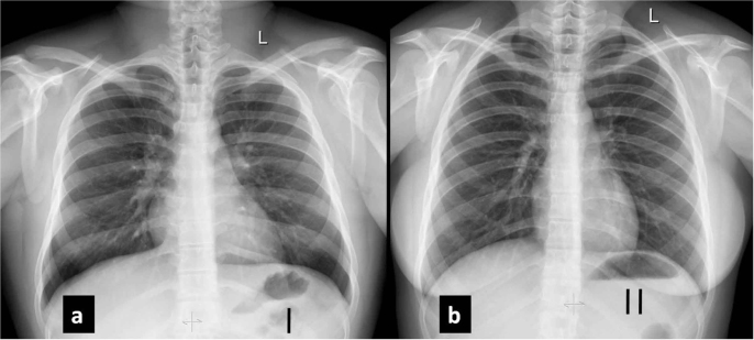
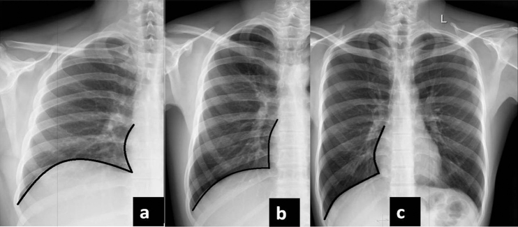
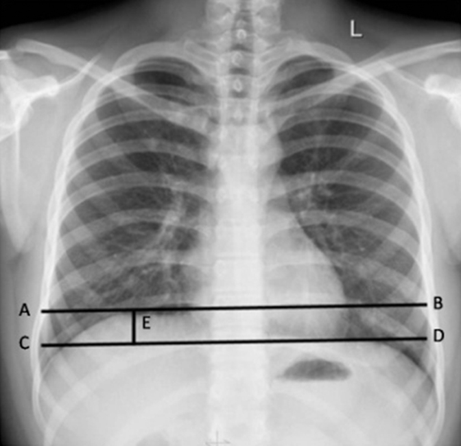
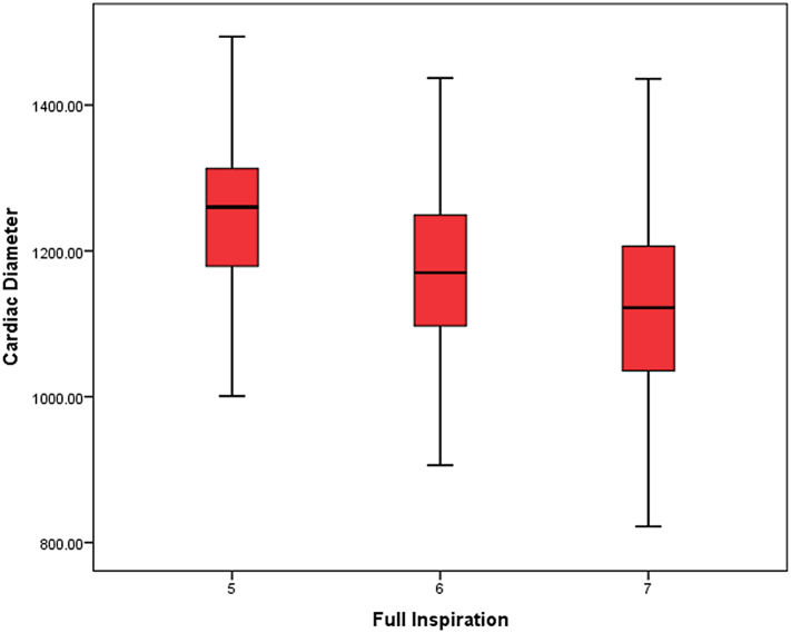
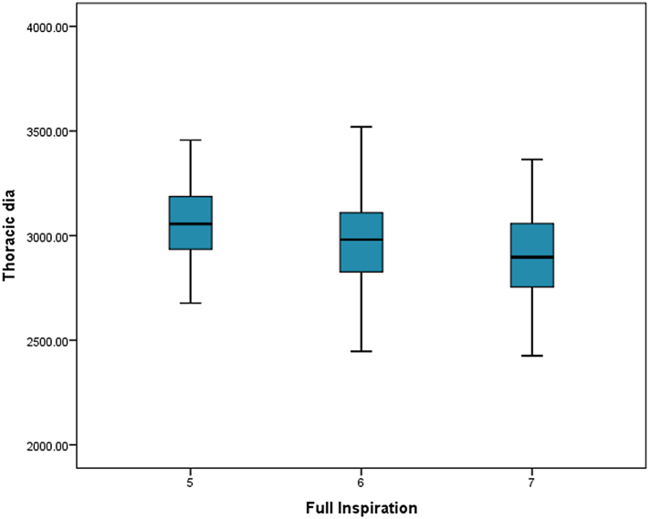
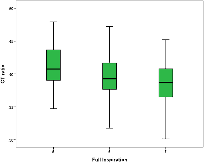
Background: Knowledge of normal variation and measurements on a chest radiograph is essential to interpret any abnormality. There is paucity of information about normal measurement ranges and variations in young adolescents, particularly from Indian subcontinent. The aim of this study was to analyze certain normal variations on screening chest radiographs of healthy Indian adolescents and the objectives were to measure/assess (1) degree of inspiration, (2) cardiothoracic ratio (CTR), (3) presence of gastric fundic bubble, (4) fundocupolic distance, (5) presence of splenic flexure, (6) difference in height of diaphragmatic domes and (7) effect of inspiration on the CTR.
Methods: Digital chest radiographs obtained during routine medical examinations for all consecutive medical graduate aspirants in the year 2016 at a medical college, were analyzed for the above mentioned parameters using DICOM viewing software.
Result: A total of 558 chest radiographs were analyzed. The mean age of the subjects was 18.50 (SD = 1.002) (range: 17-22 years). There were 497 (89.1%) male and 61 (10.9%) female. Degree of inspiration was at 5th, 6th, 7th and 8th ribs in 29 (5.1%), 259 (46.4%), 264 (47.3%) and 6 (1%) respectively. Mean maximum transverse cardiac diameter, internal thoracic diameter, CTR were 1153.22 ± 120.01, 2935.24 ± 224.86 and 0.39 ± 0.03 respectively. Females had slightly higher CTR (0.40 ± 0.035) as compared to the males (0.39 ± 0.032) (p = 0.009). Gastric fundic bubble was visualized in 91% subjects. Mean fundo-cupolic distance was 8.75 ± 8.00. Mean value for difference in the level of two domes of diaphragm was 15.28 ± 5.38.
Conclusion: The study highlights normal range of inspiration, CT ratio, fundocupolic distance and diaphragmatic dome level difference on screening chest radiographs in healthy Indian adolescents.
Keywords: Cardiothoracic ratio; Chest radiograph; Diaphragm; Fundocupolic distance; Indian adolescents.
Figures

Similar articles
-
Establishing the Cardiothoracic Ratio Using Chest Radiographs in an Indigenous Ghanaian Population: A Simple Tool for Cardiomegaly Screening.Ghana Med J. 2015 Sep;49(3):159-64. doi: 10.4314/gmj.v49i3.6. Ghana Med J. 2015. PMID: 26693191 Free PMC article.
-
Two-dimensional cardiothoracic ratio for evaluation of cardiac size in German shepherd dogs.J Vet Cardiol. 2014 Dec;16(4):237-44. doi: 10.1016/j.jvc.2014.08.001. Epub 2014 Sep 18. J Vet Cardiol. 2014. PMID: 25438928
-
Gender and Age Differences in Cardiac Size Parameters of Ghanaian Adults: Can One Parameter Fit All? Part Two.Ethiop J Health Sci. 2021 May;31(3):561-572. doi: 10.4314/ejhs.v31i3.13. Ethiop J Health Sci. 2021. PMID: 34483613 Free PMC article.
-
Changes in Cross-Sectional Area and Transverse Diameter of the Heart on Inspiratory and Expiratory Chest CT: Correlation with Changes in Lung Size and Influence on Cardiothoracic Ratio Measurement.PLoS One. 2015 Jul 7;10(7):e0131902. doi: 10.1371/journal.pone.0131902. eCollection 2015. PLoS One. 2015. PMID: 26151361 Free PMC article.
-
Radiological Cardiothoracic Ratio in Evidence-Based Medicine.J Clin Med. 2021 May 8;10(9):2016. doi: 10.3390/jcm10092016. J Clin Med. 2021. PMID: 34066783 Free PMC article. Review.
Cited by
-
Diaphragm Dysfunction After Cardiac Surgery.Braz J Cardiovasc Surg. 2025 Jun 4;40(4):e20230239. doi: 10.21470/1678-9741-2023-0239. Braz J Cardiovasc Surg. 2025. PMID: 40464402 Free PMC article.
References
-
- Dimopoulos K., Giannakoulas G., Bendayan I. Cardiothoracic ratio from postero-anterior chest radiographs: a simple, reproducible and independent marker of disease severity and outcome in adults with congenital heart disease. Int J Cardiol. 2013;166(2):453–457. - PubMed
-
- Nickol K., Wade A.J. Radiographic heart size and cardiothoracic ratio in three ethnic groups: a basis for a simple screening test for cardiac enlargement in men. Br J Radiol. 1982;55(654):399–403. - PubMed
-
- Sinha U., Sahay U.S., Athavale S.A., Depujari R., Kumar S. Comparative study of cardiac size by chest X-ray and echocardiography. J Anat Soc India. 2013;62:28–32.
LinkOut - more resources
Full Text Sources
