

Cone beam computerized tomography evaluation of incisive canal and anterior maxillary bone thickness for placement of immediate implants
- PMID: 30449964
- PMCID: PMC6180735
- DOI: 10.4103/jips.jips_167_18
Cone beam computerized tomography evaluation of incisive canal and anterior maxillary bone thickness for placement of immediate implants
Abstract
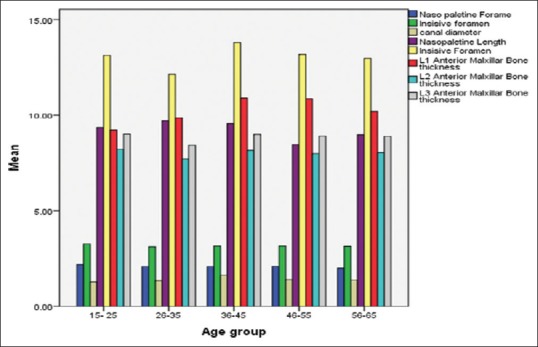
Purpose: Variation of dimensions of the nasopalatine canal and anterior maxillary bone thickness vary in relation to age, gender, edentulism, and ethnicity; thorough knowledge with regard to these landmarks is of vital importance prior to surgical procedures such as implant placement and local anesthesia in the anterior maxilla. Cone beam computerized tomography (CBCT) aids in accurate treatment planning in such situations.
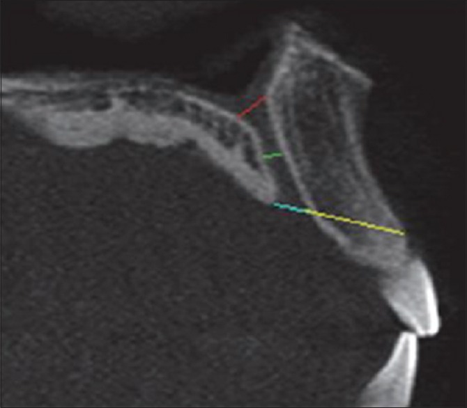
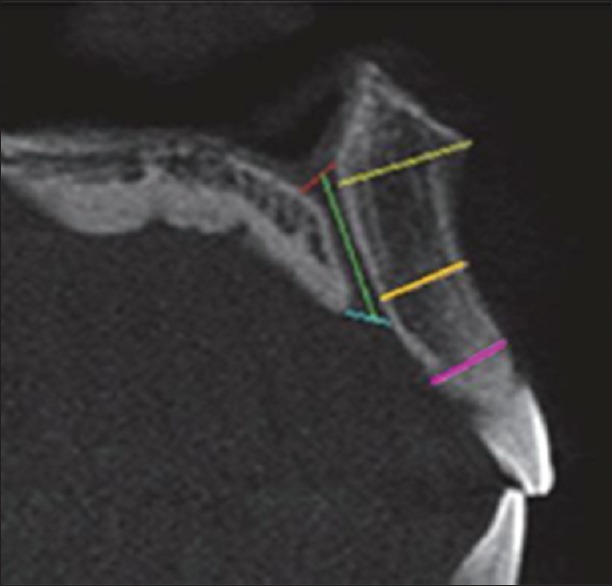
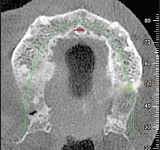
Subjects and methods: A total of 300 participants were selected by the inclusion and exclusion criteria. CBCT was performed with Hyperion X9 CBCT Scanner. Images were reconstructed from the CBCT data using NNT image reconstruction software and visualized using multi-planar resolution screen. The dimensions of the nasopalatine foramen (NPF), the incisive canal (IC) and foramen, and anterior maxillary bone thickness were measured.
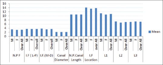
Results: The mean diameter of NPF was found to be 3.27 mm, incisive foramen (IF) was 3.62 mm, IC was 2.12 mm. The average length of the IC was 10.66 mm. The IF was located at a mean distance of 13.81 mm away from the most anteroinferior point of the cortical plate of the labial bone of the maxilla. The anterior maxillary bone was the thickest at the nasal spine level (10.94 mm), and was the narrowest at lower labial alveolus (7.16 mm). The average anterior maxillary bone thickness was found to be 8.36 mm.
Conclusion: Within the limitations of the study, it was found that found that gender and age are important factors that affected the characteristics of the IC and the amount of bone anterior to it.
Keywords: Anterior maxilla; bone thickness; cone beam computerized tomography; immediate implant; incisive canal dimensions.
Conflict of interest statement
There are no conflicts of interest.
Figures
References
-
- Vera C, De Kok IJ, Reinhold D, Limpiphipatanakorn P, Yap AK, Tyndall D, et al. Evaluation of buccal alveolar bone dimension of maxillary anterior and premolar teeth: A cone beam computed tomography investigation. Int J Oral Maxillofac Implants. 2012;27:1514–9. - PubMed
-
- Güncü GN, Yıldırım YD, Yılmaz HG, Galindo-Moreno P, Velasco-Torres M, Al-Hezaimi K, et al. Is there a gender difference in anatomic features of incisive canal and maxillary environmental bone? Clin Oral Implants Res. 2013;24:1023–6. - PubMed
LinkOut - more resources
Full Text Sources
Miscellaneous