

Three-dimensional Printing of Multilayered Tissue Engineering Scaffolds
- PMID: 30450010
- PMCID: PMC6233733
- DOI: 10.1016/j.mattod.2018.02.006
Three-dimensional Printing of Multilayered Tissue Engineering Scaffolds
Abstract
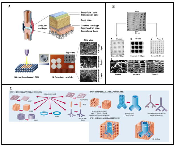
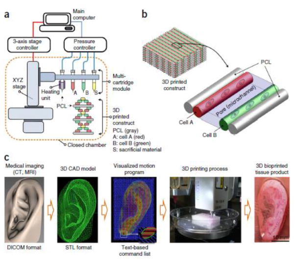
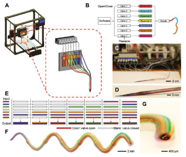
The field of tissue engineering has produced new therapies for the repair of damaged tissues and organs, utilizing biomimetic scaffolds that mirror the mechanical and biological properties of host tissue. The emergence of three-dimensional printing (3DP) technologies has enabled the fabrication of highly complex scaffolds which offer a more accurate replication of native tissue properties and architecture than previously possible. Of strong interest to tissue engineers is the construction of multilayered scaffolds that target distinct regions of complex tissues. Musculoskeletal and dental tissues in particular, such as the osteochondral unit and periodontal complex, are composed of multiple interfacing tissue types, and thus benefit from the usage of multilayered scaffold fabrication. Traditional 3DP technologies such as extrusion printing and selective laser sintering have been used for the construction of scaffolds with gradient architectures and mixed material compositions. Additionally, emerging bioprinting strategies have been used for the direct printing and spatial patterning of cells and chemical factors, capturing the complex organization found in the body. To better replicate the varied and gradated properties of larger tissues, researchers have created scaffolds composed of multiple materials spanning natural polymers, synthetic polymers, and ceramics. By utilizing high precision 3DP techniques and judicious material selection, scaffolds can thus be designed to address the regeneration of previously challenging musculoskeletal, dental, and other heterogeneous target tissues. These multilayered 3DP strategies show great promise in the future of tissue engineering.
Figures
References
-
- Fisher JP, Mikos AG, Bronzino JD, Peterson DR. Tissue Engineering: Principles and Practices [Internet] Taylor&Francis Group; 2012. [cited 2017 Jul 11]. Available from: https://www.crcpress.com/Tissue-Engineering-Principles-and-Practices/Fis....
-
- Langer R, Vacanti JP. Tissue engineering. Science. 1993 May 14;260(5110):920–6. - PubMed
-
- Bose S, Vahabzadeh S, Bandyopadhyay A. Bone tissue engineering using 3D printing. Mater Today. 2013 Dec;16(12):496–504.
-
- Sears NA, Seshadri DR, Dhavalikar PS, Cosgriff-Hernandez E. A Review of Three-Dimensional Printing in Tissue Engineering. Tissue Eng Part B Rev. 2016 Feb 9;22(4):298–310. - PubMed
-
- Shim J-H, Lee J-S, Kim JY, Cho D-W. Bioprinting of a mechanically enhanced three-dimensional dual cell-laden construct for osteochondral tissue engineering using a multi-head tissue/organ building system. J Micromechanics Microengineering. 2012;22(8):085014.
Grants and funding
LinkOut - more resources
Full Text Sources