. 2018 May 9;25(1):7100312.
doi: 10.1109/JSTQE.2018.2833205. eCollection 2019 Jan-Feb.

MAESTROS: A Multiwavelength Time-Domain NIRS System to Monitor Changes in Oxygenation and Oxidation State of Cytochrome-C-Oxidase

Affiliations

MAESTROS: A Multiwavelength Time-Domain NIRS System to Monitor Changes in Oxygenation and Oxidation State of Cytochrome-C-Oxidase

Frederic Lange et al. IEEE J Sel Top Quantum Electron. .

Abstract

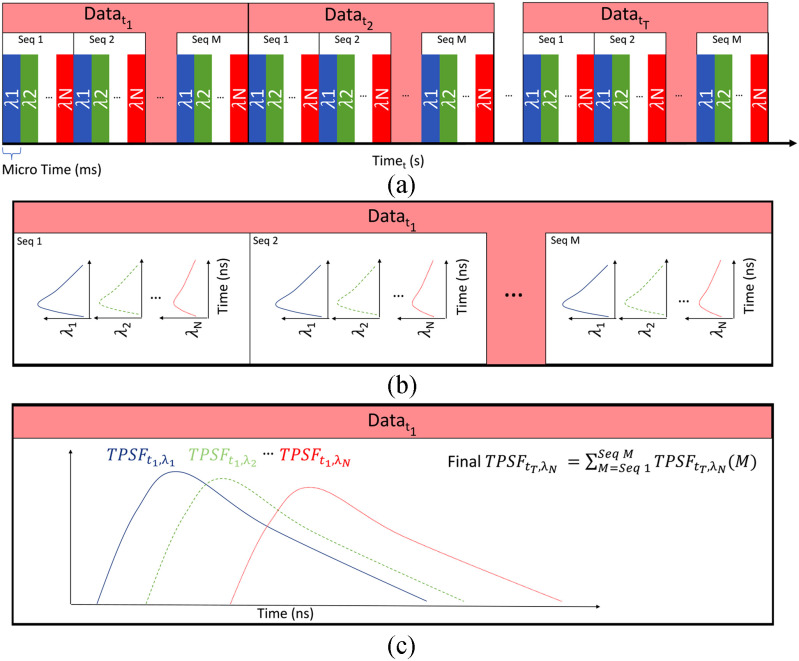
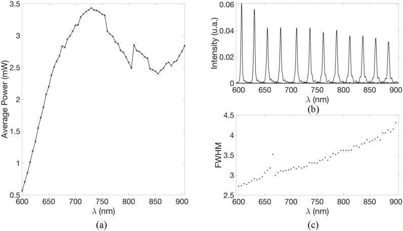
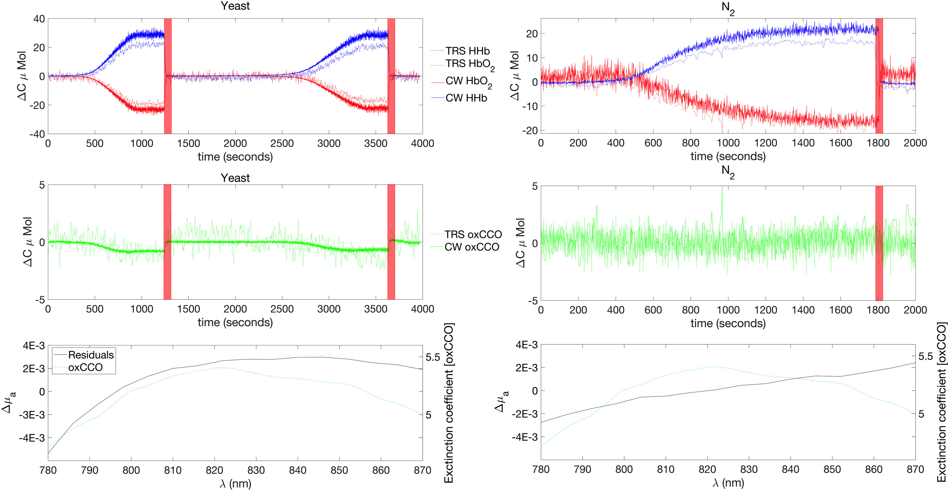
We present a multiwavelength, multichannel, time-domain near-infrared spectroscopy system named MAESTROS. This instrument can measure absorption and scattering coefficients and can quantify the concentrations of oxy- and deoxy-haemoglobin ([HbO2], [HHb]), and oxidation state of cytochrome-c-oxidase ([oxCCO]). This system is composed of a supercontinuum laser source coupled with two acousto-optic tuneable filters. The light is collected by four photomultipliers tubes, connected to a router to redirect the signal to a single time-correlated single-photon counting card. The interface between the system and the tissue is based on optical fibres. This arrangement allows us to resolve up to 16 wavelengths, within the range of 650-900 nm, at a sampling rate compatible with the physiology (from 0.5 to 2 Hz). In this paper, we describe the system and assess its performance based on two specifically designed protocols for photon migration instruments, the basic instrument protocol and nEUROPt protocols, and on a well characterized liquid phantom based on Intralipid and water. Then, the ability to resolve [HbO2 ], [HHb], and [oxCCO] is demonstrated on a homogeneous liquid phantom, based on blood for [HbO2], [HHb], and yeast for [oxCCO]. In the future, the system could be used to monitor brain tissue physiology.

Keywords: Time domain measurements; biomedical engineering; laser biomedical applications; photomultipliers; scattering; spectroscopy.


Figures

Fig. 1.
Fig. 1.
(a) Picture of the front of the system, (b) Picture of the rear of system, showing fibre optic connections. (c) Schematic illustration of the major system components.
Fig. 2.
Fig. 2.
Diagram of the acquisition sequence. (a) Diagram of the timing of the acquisition sequence. The TPSF at each wavelength is acquired sequentially with a small integration time per wavelength (10th of microseconds). Then this sequence is repeated M times. This operation is repeated for each time point. (b) Example of the acquisition of the TPSF for the first time point. This illustrate the sequential acquisition of the TPSF for each wavelength. (c) Final TPSF for the first time point. All the TPSFs of every sequence within the first time point are summed up at each wavelength and saved in the same file.
Fig. 3.
Fig. 3.
Optical arrangement of the AOTF. The input beam is first split by a beam splitter and directed to two separate AOTFs. Then the beam at the outputs of the AOTFs are focused into two separate optical fibres.
Fig. 4.
Fig. 4.
Optical properties of the VOA. (a) Optical density of the 12 positions of the VOA. (b) Spectral characteristic of position 10.
Fig. 5.
Fig. 5.
(a) Flowchart of the acquisition software. VI: Virtual instrument. (b) Typical flowchart of the data processing steps. DE: Diffusion Equation, IRF: Instrument Response Function. MBLL: Modified Beer-Lambert Law.
Fig. 6.
Fig. 6.
(a) Typical power (b) spectra and (c) FWHM of the source between 600 and 900 nm, in steps of 5 nm.
Fig. 7.
Fig. 7.
Summary of the basic characteristics of the system. (a) Responsivity of detector 2 (b) Stability of the intensity of the IRF for 16 wavelengths. Intensity expressed as the percentage of variation regarding the mean value of the last 30 minutes. (c) IRF of 16 wavelengths of detector 2 (d) Stability of the mean time of flight of the IRF for 16 wavelengths. Mean time expressed as the mean time of flight variation regarding the mean value of the last 30 minutes.
Fig. 8.
Fig. 8.
(a) Absorption spectrum of water as measured in a 1% aqueous solution of Intralipid using our system. Error bars represents the variability across all the detectors. (b) Reduced scattering spectrum of a 1% aqueous solution of Intralipid using our system. Error bars represents the variability across all the detectors.
Fig. 9.
Fig. 9.
(a) Contrast at 800 nm for detector 4, for a target of formula image, at depth between 6 mm and 30 mm, in steps of 2 mm. (b) Contrast, for detector 4, for a target of formula image, at 6 mm of depth for 2 gates (early and late) for wavelengths between 650 and 900 nm in steps of 15 mm. (c) Contrast, for detector 4, for a target of formula image, at 16 mm of depth for 2 gates (early and late) for wavelengths between 650 and 900 nm in steps of 15 mm. Error bars represent noise as obtained from the standard deviation of 100 repeated measurements.
Fig. 10.
Fig. 10.
Schematic of the phantom measurement configuration. Optical fibres are not shown for clarity.
Fig. 11.
Fig. 11.
(a) evolution of the saturation of the yeast (solid black) and formula image (dashed grey) phantoms over the first hour of the experiment. (b) Example of the fitting of the optical properties for the formula image phantom at 792 nm. The blue curve represents a low saturation point and the red curve represents a high saturation point. The exact timing of those points is reported with stars on part (a).
Fig. 12.
Fig. 12.
Summary of the phantom results. From top to bottom. Concentration changes in [HHb] and [HbO2] for both the TR and CW instrument, Concentration changes in [oxCCO] for both the TR and CW instrument, Residuals of the 2 and 3 components fit together with the extinction coefficient of the [oxCCO]. Left and right columns refer to yeast and formula image phantom respectively. The red shaded regions correspond to the O2 ON period.
Fig. 13.
Fig. 13.
Concentration changes for [HHb], [HbO2] and [oxCCO] consecutive to a muscular cuff occlusion. Reproduced from reference .

References

    1. Ferrari M. and Quaresima V. , “A brief review on the history of human functional near-infrared spectroscopy (fNIRS) development and fields of application,” Neuroimage, vol. 63 , no. 2, pp. 921–935, Nov. 2012. - PubMed
    1. Quaresima V. and Ferrari M., “Functional near-infrared spectroscopy (fNIRS) for assessing cerebral cortex function during human behavior in natural/social situations: A concise review,” Organ. Res. Methods , 1094428116658959, pp. 1–23, Jul. 2016.
    1. Ferrari M., Mottola L., and Quaresima V., “ Principles, techniques, and limitations of near infrared spectroscopy,” Can. J. Appl. Physiol., vol. 29, no. 4, pp. 463– 487, 2004. - PubMed
    1. Pellicer A. and Bravo M. D. C., “Near-infrared spectroscopy: a methodology-focused review,” Semin. Fetal Neonatal Med., vol. 16, no. 1, pp. 42–9, Feb. 2011. - PubMed
    1. McLellan S. and Walsh T., “Oxygen delivery and haemoglobin,” Contin. Edu. Anaesthesia Crit. Care Pain, vol. 4, no. 4, pp. 123– 126, Aug. 2004.

LinkOut - more resources