Review
. 2018 Nov 2:9:2541.
doi: 10.3389/fimmu.2018.02541. eCollection 2018.

Interplay Between the Unfolded Protein Response and Immune Function in the Development of Neurodegenerative Diseases

Affiliations
Review

Interplay Between the Unfolded Protein Response and Immune Function in the Development of Neurodegenerative Diseases

Paulina García-González et al. Front Immunol. .

Abstract

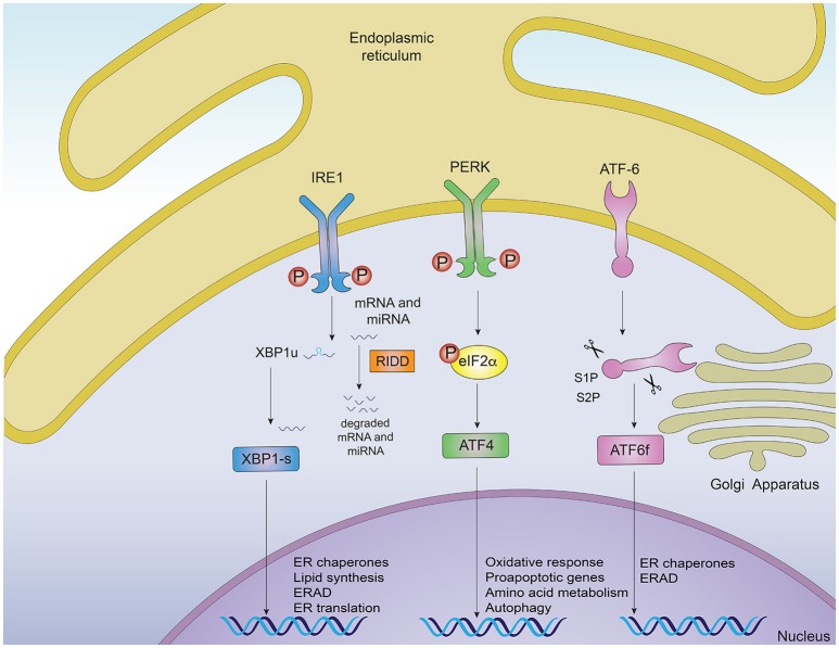
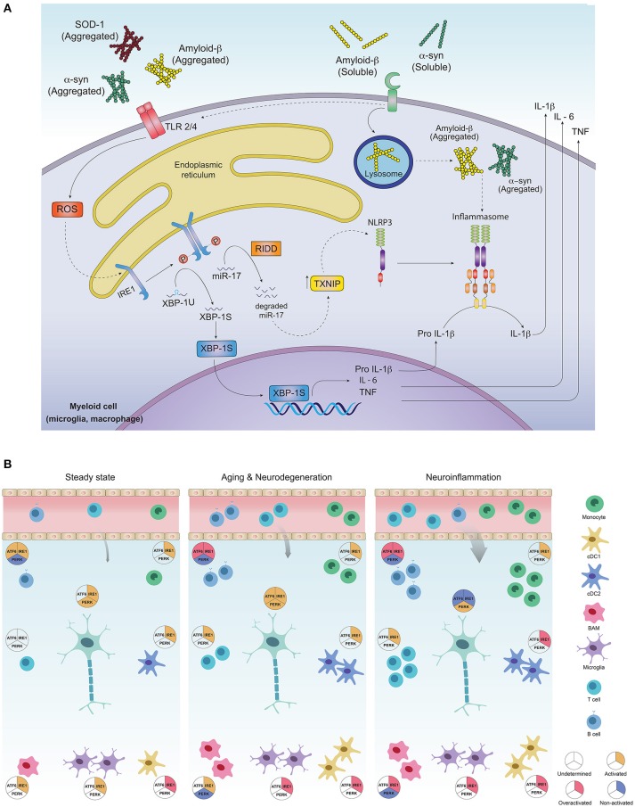
Emerging evidence suggests that the immune and nervous systems are in close interaction in health and disease conditions. Protein aggregation and proteostasis dysfunction at the level of the endoplasmic reticulum (ER) are central contributors to neurodegenerative diseases. The unfolded protein response (UPR) is the main transduction pathway that maintains protein homeostasis under conditions of protein misfolding and aggregation. Brain inflammation often coexists with the degenerative process in different brain diseases. Interestingly, besides its well-described role in neuronal fitness, the UPR has also emerged as a key regulator of ontogeny and function of several immune cell types. Nevertheless, the contribution of the UPR to brain inflammation initiated by immune cells remains largely unexplored. In this review, we provide a perspective on the potential role of ER stress signaling in brain-associated immune cells and the possible implications to neuroinflammation and development of neurodegenerative diseases.

Keywords: ER stress; UPR; immune cells; immune system; inflammation; misfolded proteins; neurodegeneration; protein protein misfolding diseases.


Figures

Figure 1
Figure 1
Signaling pathways of the unfolded protein response. Noxious stimuli in cells may induce endoplasmic reticulum (ER) stress and trigger an adaptive response known as the unfolded protein response (UPR), which is controlled by three main ER-resident sensors: IRE1α, PERK and ATF6. Upon ER stress, IRE1α autophosphorylates, leading to the activation of its RNase domain and the processing of the mRNA encoding for XBP1s, a transcriptional factor that upregulates genes involved in protein folding and quality control, in addition to regulating ER/Golgi biogenesis and ER-mediated degradation (ERAD). Additionally, IRE1α RNase also degrades a subset of specific RNAs and microRNAs, a process termed Regulated IRE1α-Dependent Decay (RIDD). The second ER sensor, PERK, phosphorylates the translation of the eukaryotic initiation factor eIF2α, decreasing the synthesis of proteins and the overload of misfolded proteins at the ER. PERK phosphorylation also leads to the specific translation of ATF4, a transcription factor that promotes the expression of genes related to amino acid metabolism, anti-oxidant response, autophagy and apoptosis. The third UPR sensor, ATF6, is a type II ER transmembrane protein that encodes a bZIP transcriptional factor in its cytosolic domain. Following ER stress, ATF6 translocates to the Golgi apparatus where it is processed, releasing a transcription factor which directs the expression of genes encoding ER chaperones, ERAD components and molecules involved in lipid biogenesis.
Figure 2
Figure 2
Activation of the unfolded protein response in CNS-residing immune cells may contribute to neuroinflammation and PMDs development. (A) Protein aggregates can promote inflammation via triggering of innate receptors and activation of the UPR. Neurodegeneration associated molecular patterns (NAMPS) such as protein aggregates are recognized by pattern recognition receptors (TLRs and PRRs) present on immune cells and signal through ROS production, which in turn could activate the IRE1α/XBP1s axis for co-transcriptional activation of IL-6, TNF and IL-1β. On the other hand, through RIDD, IRE1α induces degradation of the TXNIP-destabilizing microRNA mir-17, allowing activation of the NLRP3 inflammasome and processing of IL-1β into its active form. (B) Most of the immune cell lineages residing in the healthy and pathogenic brain are known targets of the UPR in peripheral tissues. In steady state, the most abundant immune cells in the brain are microglia, which along with border associated macrophages (“BAMs”) and dendritic cells act as sentinels, sampling the environment and clearing cell debris, maintaining CNS homeostasis. Except for dendritic cells and macrophages, which exhibit IRE1α/XBP1s activation, little is known about UPR activation in additional myeloid subsets, although microglia, macrophages and monocytes could potentially activate this axis downstream of PRR signaling. While very rare, B and T cells have been identified in the steady state brain, and activation of IRE1α/XBP1s has been proposed to be critical for their differentiation and activation. ATF6 axis is also necessary for B cell development and activation whilst absence of PERK contributes to plasma cell differentiation and immunoglobulin synthesis. Basal activation of UPR in neurons is still a matter of debate in literature as the function of IRE1α and PERK pathways has just begun to be understood in the context of normal neuronal physiology (72). In aging and neurodegeneration, the number of immune cells within the brain increases, due to higher cell activation as well as blood brain barrier infiltrates. Extracellular protein aggregation promotes activation of immune cells via PRRs, in addition to inducing ER stress and activation of the UPR, mainly the IRE1α/XBP1s axis. Microglia and dendritic cells become more activated, with higher production of pro-inflammatory and oxidative mediators and loss of their protein clearance function. This is further aggravated by antibodies against CNS-derived antigens by B cells accumulated in the CSF, mediated by the activation of IRE1α and ATF6 signaling. Activation of infiltrating T cells reactive to α-synuclein, amyloid-β and myelin constituents further amplify inflammation, resulting in more protein aggregation and neuronal loss. In neurons, UPR triggering may elicit both, adaptive or neurodegenerative responses, since all three UPR pathways are engaged in brain diseases and have been found to be altered during the normal aging process. Different inducers of neuroinflammation, have shown to engage the UPR in neurons and promote a greater inflammatory response due to immune cell infiltration, mainly B and T cells. The cDC1 subset of dendritic cells could activate IRE1α for cross presentation of antigens to infiltrating CD8+ T cells, and cDC2 as well as monocyte-derived DCs may set an inflammatory environment through cytokine secretion and activation of infiltrating CD4+ T cells. Macrophages and microglia also become highly activated and could tune IRE1α/XBP1s upon recognition of NAMPs. Inflammatory mediators such as cytokines prime axonal destruction and neuronal loss. It remains to be addressed weather UPR triggering in these cells corresponds to a homeostatic (adaptive) response, or a terminal (neurodegenerative) response due to sustained unresolved ER stress.

Similar articles

Cited by

References

    1. Balch WE, Morimoto RI, Dillin A, Kelly JW. Adapting proteostasis for disease intervention. Science (2008) 319:916–9. 10.1126/science.1141448 - DOI - PubMed
    1. Tavernier SJ, Lambrecht BN, Janssens S. The unfolded protein response in the immune cell development: putting the caretaker in the driving seat. Curr Top Microbiol Immunol. (2018) 414:45–72. 10.1007/82_2017_1 - DOI - PubMed
    1. Taylor RC, Berendzen KM, Dillin A. Systemic stress signalling: understanding the cell non-autonomous control of proteostasis. Nat Rev Mol Cell Biol.(2014) 15:211. 10.1038/nrm3752 - DOI - PMC - PubMed
    1. Cabral-Miranda F, Hetz C. ER Stress and neurodegenerative disease: a cause or effect relationship? Curr Top Microbiol Immunol. (2018) 414:131–57. 10.1007/82_2017_52 - DOI - PubMed
    1. Soto C. Unfolding the role of protein misfolding in neurodegenerative diseases. Nat Rev Neurosci. (2003) 4:49–60. 10.1038/nrn1007 - DOI - PubMed

Publication types