

X-linked juvenile retinoschisis: phenotypic and genetic characterization
- PMID: 30450322
- PMCID: PMC6232335
- DOI: 10.18240/ijo.2018.11.22
X-linked juvenile retinoschisis: phenotypic and genetic characterization
Abstract
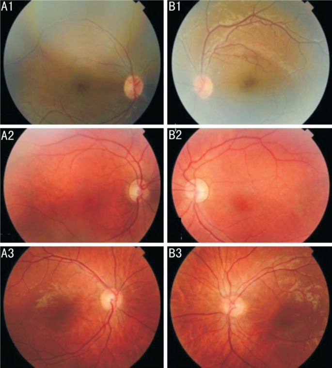
Juvenile X-linked retinoschisis (XLRS, MIM#312700) belongs to a group of the vitreoretinal dystrophies. We aimed to describe the phenotype-genotype correlation of three XLRS cases in juveniles with different novel mutations from the Lithuanian population. The patients demonstrated macular retinoschisis and typical cyst-like cavities on spectral-domain optical coherence tomography (SD-OCT) images. The mean central foveal thickness was 569.7 µm. Two patients presented with peripheral retinoschisis. Flash electroretinogram demonstrated a reduced b/a ratio (<1.0) in all patients. RS1 (NM_000330.3) gene coding exons Sanger sequencing was performed. RS1 c.599G>T (p.R200L) mutation was detected in one case, showing to be pathogenic in silico analysis. c. (92_97) insC (p.W33fs) mutation was identified for another patient, indicating the variant is possibly damaging in silico analysis. The third case was identified with a pathogenic mutation c.422C>G (p.R141H), HGMD CM981753. These are the first cases of XLRS in the Lithuanian population confirmed by molecular genotyping. Presented patients had a different genotype but similar phenotypic traits.
Keywords: RS1 mutation; X-linked retinoschisis; electroretinogram; optical coherence tomography.
Figures

References
-
- Riveiro-Alvarez R, Trujillo-Tiebas MJ, Gimenez-Pardo A, Garcia-Hoyos M, Lopez-Martinez MA, Aguirre-Lamban J, Garcia-Sandoval B, Vazquez-Fernandez del Pozo S, Cantalapiedra D, Avila-Fernandez A, Baiget M, Ramos C, Ayuso C. Correlation of genetic and clinical findings in Spanish patients with X-linked juvenile retinoschisis. Invest Ophthalmol Vis Sci. 2009;50(9):4342–4350. - PubMed
LinkOut - more resources
Full Text Sources