. 2019 Jan;23(1):487-496.
doi: 10.1111/jcmm.13951. Epub 2018 Nov 18.

Efficient method for isolation of reticulocyte RNA from healthy individuals and hemolytic anaemia patients

Affiliations

Efficient method for isolation of reticulocyte RNA from healthy individuals and hemolytic anaemia patients

Michał Skulski et al. J Cell Mol Med. 2019 Jan.

Abstract

Despite enormous progress and development of high-throughput methods in genome-wide mRNA analyses, data on the erythroid transcriptome are still limited, even though they could be useful in medical diagnostics and personalized therapy as well as in research on normal and pathological erythroid maturation. Although obtaining normal and pathological reticulocyte transcriptome profiles should contribute greatly to our understanding of the molecular bases of terminal erythroid differentiation as well as the mechanisms of the hematological diseases, a basic limitation of these studies is the difficulty of efficient reticulocyte RNA isolation from human peripheral blood. The restricted number of possible parallel experiments primarily concern healthy individuals with the lowest number of reticulocytes in the peripheral blood and a low RNA content. In the present study, an efficient method for reticulocyte RNA isolation from healthy individuals and hemolytic anaemia patients is presented. The procedure includes leukofiltration, Ficoll-Paque gradient centrifugation, Percoll gradient centrifugation, and negative (CD45 and CD61) immunomagnetic separation. This relatively fast and simple four-stage method was successfully applied to obtain a reticulocyte-rich population from healthy subjects, which was used to efficiently isolate the high-quality RNA essential for successful NGS-based transcriptome analysis.

Keywords: RNA-Seq; hereditary hemolytic anaemia; hereditary spherocytosis; reticulocyte isolation; reticulocyte transcriptome.


Figures

Figure 1
Figure 1
Scheme of four‐step purification procedure used to obtain reticulocyte‐rich suspension from human peripheral blood
Figure 2
Figure 2
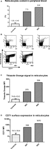
Reticulocyte characteristics. A, Examples of reticulocyte quantification in human peripheral blood. Examination was performed for healthy individuals (C1‐C3) and for hemolytic anaemia patients (N61 & N62). The number of reticulocytes was determined from the dot plots: FSC (forward scatter) vs TO fluorescence. B, Analysis of CD71 surface exposure in WBC and platelet‐free reticulocyte preparations obtained from the healthy individuals and HA patients allows determination of reticulocyte number and maturity status. Dot plots showing CD71 surface expression and RNA content (TO) in reticulocytes isolated from healthy individuals and HA patients (N61, N62). These data indicate a significantly higher number of immature (early) reticulocytes in peripheral blood of HA patients than in healthy individuals. C, Quantification of purified reticulocyte measured by Thiazole Orange (TO, RNA content) fluorescence reveals 3.6 and 6.7 times higher signals for N61 and N62 HA patients, respectively, than in healthy individuals (controls: n = 3). D, CD71 surface expression in purified reticulocytes for N61 and N62 HA patients is significant
Figure 3
Figure 3
RNA amounts for each examined group. Healthy individuals (n = 4), C14—hereditary spherocytosis patient and HA—hemolytic anaemia patients (n = 2)
Figure 4
Figure 4
Example of reticulocyte cDNA quality control. Agarose gel electrophoresis of RTPCR products (image inverted, black/white) obtained using reticulocyte cDNA from patients (HA patient, N62, and HS patient, C14) and controls (healthy individuals: C1 and C2) and primers encoding sequences of the following genes: CD45 (PTPRC gene; 217 bp)—leukocyte marker; β‐globin (HBB gene; 397 bp)—erythroid marker; integrin‐β3 (ITGB3 gene; 198 bp)—platelet marker. As the “low abundance” gene transcript PPARA (peroxisome proliferator activated receptor alpha; 324 bp) was chosen and was taken from the end of the reticulocyte cDNA library (NCBI, Library 11923). For the loading control β‐actin primers (ACTB gene; 479 bp) were used. All primer sequences used in this experiment are included in Supporting Information Table S2. As a standard “GeneRuler 100 bp DNA Ladder” (Thermo Fisher Scientific, Waltham, MA, USA) was used. Results show no leukocyte or thrombocyte RNA contamination for all analysed cases. The lowest band corresponds to primers
Figure 5
Figure 5
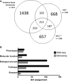
RNA‐Seq analysis. A, Comparison of the number of gene transcripts identified by our RNA‐Seq approach with the ones deposited in libraries representing the early (CD71++++) and late (GPA ++) reticulocyte maturation stages. Almost 35% of all identified gene transcripts in reticulocytes are identical to these deposited in libraries and approximately 30% of gene transcripts in both libraries are the same. Two hundred and eleven gene transcripts are common to the three groups discussed. Only transcripts successfully identified in all four individual RNA‐Seq analyses were considered. A detailed gene transcript list is provided in Supporting Information Table S6. Compare to Supporting Information Table S5. B, GeneAnalytics results. Comparison of reticulocyte transcriptome profiles obtained from RNA microarrays results previously published by Goh et al21 and our RNA‐Seq results placed in a physiological context was generated using the GeneAnalytics web server

References

    1. Gronowicz G, Swift H, Steck TL. Maturation of the reticulocyte in vitro. J Cell Sci. 1984;71:177‐197. - PubMed
    1. Lee E, Choi HS, Hwang JH, Hoh JK, Cho YH, Baek EJ. The RNA in reticulocytes is not just debris: it is necessary for the final stages of erythrocyte formation. Blood Cells Mol Dis. 2014;53:1‐10. - PubMed
    1. Hu J, Liu J, Xue F, et al. Isolation and functional characterization of human erythroblasts at distinct stages: implications for understanding of normal and disordered erythropoiesis in vivo. Blood. 2013;121:3246‐3253. - PMC - PubMed
    1. Ney PA. Normal and disordered reticulocyte maturation. Curr Opin Hematol. 2011;18:152‐157. - PMC - PubMed
    1. d'Onofrio G, Kuse R, Foures C, Jou JM, Pradella M, Zini G. Reticulocytes in haematological disorders. Clin Lab Haematol. 1996;18(Suppl 1):29‐34. - PubMed

Publication types