Osterix promotes the migration and angiogenesis of breast cancer by upregulation of S100A4 expression
- PMID: 30450809
- PMCID: PMC6349213
- DOI: 10.1111/jcmm.14012
Osterix promotes the migration and angiogenesis of breast cancer by upregulation of S100A4 expression
Abstract
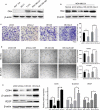
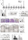
As a key transcription factor required for bone formation, osterix (OSX) has been reported to be overexpressed in various cancers, however, its roles in breast cancer progression remain poorly understood. In this study, we demonstrated that OSX was highly expressed in metastatic breast cancer cells. Moreover, it could upregulate the expression of S100 calcium binding protein A4 (S100A4) and potentiate breast cancer cell migration and tumor angiogenesis in vitro and in vivo. Importantly, inhibition of S100A4 impaired OSX-induced cell migration and capillary-like tube formation. Restored S100A4 expression rescued OSX-short hairpin RNA-suppressed cell migration and capillary-like tube formation. Moreover, the expression levels of OSX and S100A4 correlated significantly in human breast tumors. Our study suggested that OSX acts as an oncogenic driver in cell migration and tumor angiogenesis, and may serve as a potential therapeutic target for human breast cancer treatment.
Keywords: S100A4; angiogenesis; breast cancer; migration; osterix.
© 2018 The Authors. Journal of Cellular and Molecular Medicine published by John Wiley & Sons Ltd and Foundation for Cellular and Molecular Medicine.
Figures


Similar articles
-
S100A4 promotes invasion and angiogenesis in breast cancer MDA-MB-231 cells by upregulating matrix metalloproteinase-13.Acta Biochim Pol. 2012;59(4):593-8. Epub 2012 Nov 16. Acta Biochim Pol. 2012. PMID: 23162804
-
Invasion and increased expression of S100A4 and CYR61 in mesenchymal transformed breast cancer cells is downregulated by GnRH.Int J Oncol. 2016 Jun;48(6):2713-21. doi: 10.3892/ijo.2016.3491. Epub 2016 Apr 18. Int J Oncol. 2016. PMID: 27098123
-
S100A4 expression is closely linked to genesis and progression of glioma by regulating proliferation, apoptosis, migration and invasion.Asian Pac J Cancer Prev. 2015;16(7):2883-7. doi: 10.7314/apjcp.2015.16.7.2883. Asian Pac J Cancer Prev. 2015. PMID: 25854377
-
Estrogen‑related receptor γ promotes the migration and metastasis of endometrial cancer cells by targeting S100A4.Oncol Rep. 2018 Aug;40(2):823-832. doi: 10.3892/or.2018.6471. Epub 2018 Jun 4. Oncol Rep. 2018. PMID: 29901152
-
The Multifaceted S100A4 Protein in Cancer and Inflammation.Methods Mol Biol. 2019;1929:339-365. doi: 10.1007/978-1-4939-9030-6_22. Methods Mol Biol. 2019. PMID: 30710284 Review.
Cited by
-
Single-Cell Transcriptomics Reveals the Complexity of the Tumor Microenvironment of Treatment-Naive Osteosarcoma.Front Oncol. 2021 Jul 21;11:709210. doi: 10.3389/fonc.2021.709210. eCollection 2021. Front Oncol. 2021. PMID: 34367994 Free PMC article.
-
Quantitative shear wave elastography in primary invasive breast cancers, based on collagen-S100A4 pathology, indicates axillary lymph node metastasis.Quant Imaging Med Surg. 2020 Mar;10(3):624-633. doi: 10.21037/qims.2020.02.18. Quant Imaging Med Surg. 2020. PMID: 32269923 Free PMC article.
-
Identification of novel prognostic biomarkers for osteosarcoma: a bioinformatics analysis of differentially expressed genes in the mesenchymal stem cells from single-cell sequencing data set.Transl Cancer Res. 2022 Oct;11(10):3841-3852. doi: 10.21037/tcr-22-2370. Transl Cancer Res. 2022. PMID: 36388032 Free PMC article.
-
SLAMF8 regulates osteogenesis and adipogenesis of bone marrow mesenchymal stem cells via S100A6/Wnt/β-catenin signaling pathway.Stem Cell Res Ther. 2024 Oct 8;15(1):349. doi: 10.1186/s13287-024-03964-1. Stem Cell Res Ther. 2024. PMID: 39380096 Free PMC article.
-
Ca2+ Signaling as the Untact Mode during Signaling in Metastatic Breast Cancer.Cancers (Basel). 2021 Mar 23;13(6):1473. doi: 10.3390/cancers13061473. Cancers (Basel). 2021. PMID: 33806911 Free PMC article. Review.
References
-
- Chen W, Zheng R, Baade PD, et al. Cancer statistics in China, 2015. CA Cancer J Clin. 2015;66:115‐132. - PubMed
-
- Siegel RL, Miller KD, Jemal A. Cancer statistics, 2016. CA Cancer J Clin. 2016;66:7‐30. - PubMed
-
- Steeg PS. Tumor metastasis: mechanistic insights and clinical challenges. Nat Med. 2006;12:895‐904. - PubMed
-
- Nakashima K, Zhou X, Kunkel G, et al. The novel zinc finger‐containing transcription factor osterix is required for osteoblast differentiation and bone formation. Cell. 2002;108:17‐29. - PubMed
-
- Logothetis CJ, Lin SH. Osteoblasts in prostate cancer metastasis to bone. Nat Rev Cancer. 2005;5:21‐28. - PubMed
Publication types
MeSH terms
Substances
LinkOut - more resources
Full Text Sources
Other Literature Sources
Medical
Research Materials

