Review
. 2018 Dec;66(12):1716-1726.
doi: 10.4103/ijo.IJO_893_18.

Choroidal biomarkers

Affiliations
Review

Choroidal biomarkers

Francesco Pichi et al. Indian J Ophthalmol. 2018 Dec.

Abstract

A structurally and functionally intact choroid tissue is vitally important for the retina function. Although central retinal artery is responsible to supply the inner retina, choroidal vein network is responsible for the remaining one-third of the external part. Abnormal choroidal blood flow leads to photoreceptor dysfunction and photoreceptor death in the retina, and the choroid has vital roles in the pathophysiology of many diseases such as central serous chorioretinopathy, age-related macular degeneration, pathologic myopia, Vogt-Koyanagi-Harada disease. Biomarkers of choroidal diseases can be identified in various imaging modalities that visualize the choroid. Indocyanine green angiography enables the visualization of choroid veins under the retinal pigment epithelium and choroidal blood flow. New insights into a precise structural and functional analysis of the choroid have been possible, thanks to recent progress in retinal imaging based on enhanced depth imaging (EDI) and swept-source optical coherence tomography (SS-OCT) technologies. Long-wavelength SS-OCT enables the choroid and the choroid-sclera interface to be imaged at greater depth and to quantify choroidal thickness profiles throughout a volume scan, thus exposing the morphology of intermediate and large choroidal vessels. Finally, OCT angiography allows a dye-free evaluation of the blood flow in the choriocapillaris and in the choroid. We hereby review different imaging findings of choroidal diseases that can be used as biomarkers of activity and response to the treatment.

Keywords: Choriocapillaris; choroid; enhanced depth imaging optical coherence tomography; indocyanine green angiography; optical coherence tomography angiography; swept-source optical coherence tomography.


Conflict of interest statement

There are no conflicts of interest

Figures

Figure 1
Figure 1
Atrophy of the outer retina bands and the RPE allows an “artifact-free” visualization of the choroid on optical coherence tomography angiography. Optical coherence tomography angiography illustrates that the choriocapillaris is characterized by a granular pattern of bright and dark (signal voids) areas (a), and visualizes the larger choroidal vessels and their anastomosis (b)
Figure 2
Figure 2
Schematic representation of the vascularization of the choroid. Perfusion of the choroid comes from the posterior ciliary arteries and from the perforating anterior ciliary arteries, while venous blood drains through the vortex system
Figure 3
Figure 3
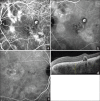
The gold standard to assess choroidal hyperpermeability in central serous chorioretinopathy remains indocyanine green angiography (a). A less invasive method is represented by optical coherence tomography angiography of the choriocapillaris that shows (with a moderate correspondence with the indocyanine green angiography leak) areas of flow void surrounded by hyperintense areas of increase flow (b): There seems to be a reactive hyperperfusion that could cause an increased hydrostatic pressure within the fenestrated vessels of the choriocapillaris. Enhanced-depth imaging optical coherence tomography (c) shows an increased thickness of the whole choroid
Figure 4
Figure 4
Choroidal hyperpermeability demonstrated on indocyanine green angiography in the both eyes of a patient presenting with polypoidal choroidal vasculopathy in the left eye. (a) Early phase (1 min) indocyanine green angiography in which branching vascular network and polyps are seen. In addition, dilated large choroidal vessels can be seen temporal to the optic disc. (b) Mid-phase (5 min) indocyanine green angiography in which diffuse hyperfl uoresecence can be seen in areas with pachyvessels. (c) Choroidal hyperpermeability was also evident from the mid phase indocyanine green angiography of the fellow eye in this patient. (d) Markedly thickened choroid (>500 μm) can be detected in the left eye using spectral-domain optical coherence tomography
Figure 5
Figure 5
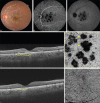
Multimodal imaging of a young female patient with tubercular serpiginous like choroiditis. Fundus photograph shows yellowish serpiginous like choroiditis lesions in the macula (a). Indocyanine green angiography shows early (b) and late (c) hypocyanescence suggestive of choriocapillaris ischemia. Enhanced-depth imaging optical coherence tomography shows choriocapillaris thickening and hypo-reflectivity corresponding to the serpiginous-like choroiditis lesions (yellow dotted line) (d). Optical coherence tomography angiography en face image at the level of choriocapillaris shows dark flow deficit areas (yellow arrowheads), which correspond to the hypocyanescent lesions on indocyanine green angiography and choriocapillaris thickening on enhanced-depth imaging optical coherence tomography (e). In the healed stage, enhanced-depth imaging optical coherence tomography shows choriocapillaris thinning (yellow dotted line) and increased choroidal reflectance (f). Optical coherence tomography angiography shows resolution of the dark areas of flow deficit
Figure 6
Figure 6
Differentiation between choriocapillaris ischemia and choriocapillaris atrophy in a patient of serpiginous like choroiditis on indocyanine green angiography and optical coherence tomography angiography imaging. In active serpiginous-like choroiditis, early phase indocyanine green angiography shows dark hypocyanescent lesions with fuzzy margins suggestive of choriocapillaris ischemia (a). In the healed stage of serpiginous like choroiditis, indocyanine green angiography shows hypocyanescent lesions with less fuzzy borders and visibility of ill-defined underlying choroidal vessels suggestive of choriocapillaris atrophy (b). Optical coherence tomography angiography image of the acute stage lesions shows dark areas of flow deficit corresponding to the areas of choriocapillaris ischemia on indocyanine green angiography (c). In the healed stage, there is resolution of areas of flow deficit with visibility of underlying medium sized choroidal vessels in areas of choriocapillaris atrophy (d)
Figure 7
Figure 7
Multimodal imaging in a 29-year-old male patient of Vogt–Koyanagi–Harada disease. Fundus photograph of the left eye shows presence of exudative retinal detachment involving the macula with underlying creamy yellowish-white choroiditis lesions (a). Indocyanine green angiography shows multiple hypocyanescent lesions in the early phase (b) which remain hypocyanescent or isocyanescent in the late phase (c). Optical coherence tomography angiography en face image at the level of the choriocapillaris shows multiple dark areas of flow deficit (yellow arrowheads) suggestive of choriocapillaris ischemia (d). The structural en face scan does not show loss of signal transmission (e). Swept-source optical coherence tomography shows increase in total choroidal thickness and clear demarcation of the choroidoscleral interface (white arrowheads) (f). Optical coherence tomography angiography at 3-month follow-up shows resolution of most areas of flow deficit indicating resolution of choriocapillaris ischemia with minimal choriocapillaris atrophy (g). The corresponding structural en face scan shows no loss of signal transmission (h). Swept-source optical coherence tomography at 3-month follow-up visit shows marked reduction in the total choroidal thickness (i)

References

    1. Hogan MJ, Alvarado JA, Wedell JE. Histology of the Human Eye. Philadelphia: W.B. Saunders; 1971.
    1. De Stefano ME, Mugnaini E. Fine structure of the choroidal coat of the avian eye. Vascularization, supporting tissue and innervation. Anat Embryol (Berl) 1997;195:393–418. - PubMed
    1. Olver JM. Functional anatomy of the choroidal circulation: Methyl methacrylate casting of human choroid. Eye (Lond) 1990;4(Pt 2):262–72. - PubMed
    1. Spraul CW, Lang GE, Lang GK, Grossniklaus HE. Morphometric changes of the choriocapillaris and the choroidal vasculature in eyes with advanced glaucomatous changes. Vision Res. 2002;42:923–32. - PubMed
    1. Krebs W, Krebs IP. Ultrastructural evidence for lymphatic capillaries in the primate choroid. Arch Ophthalmol. 1988;106:1615–6. - PubMed

MeSH terms