. 2019 Mar;43(3):314-324.
doi: 10.1097/PAS.0000000000001167.

Morphologic Overlap Between Inflammatory Myofibroblastic Tumor and IgG4-related Disease: Lessons From Next-generation Sequencing

Affiliations

Morphologic Overlap Between Inflammatory Myofibroblastic Tumor and IgG4-related Disease: Lessons From Next-generation Sequencing

Martin S Taylor et al. Am J Surg Pathol. 2019 Mar.

Abstract

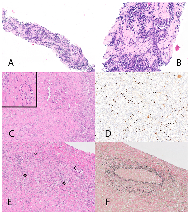
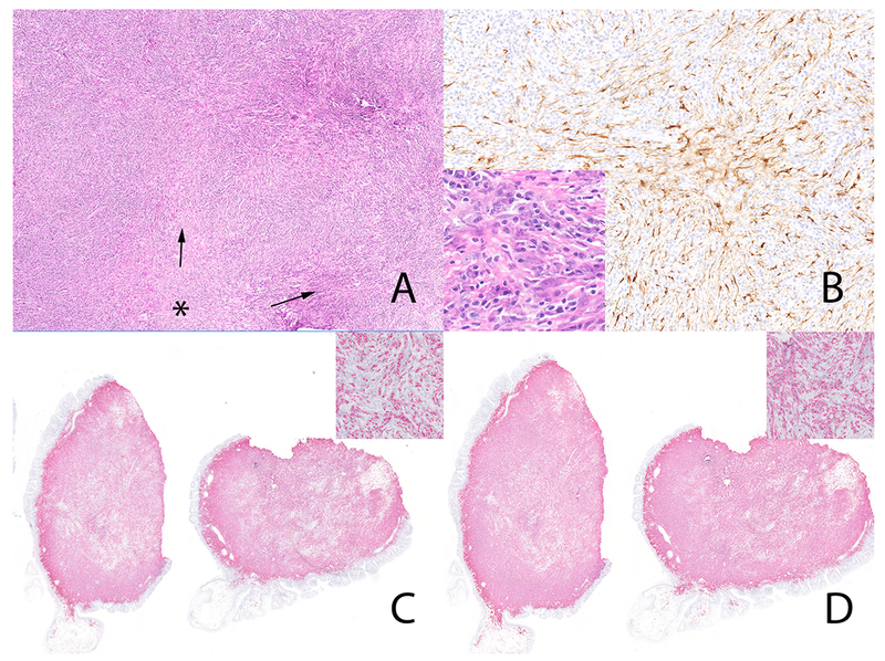
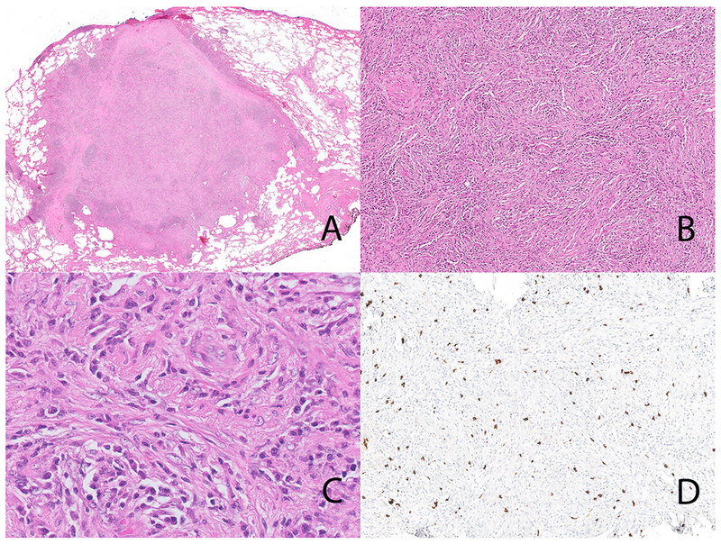
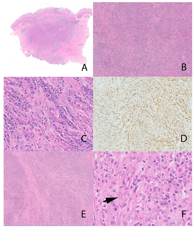
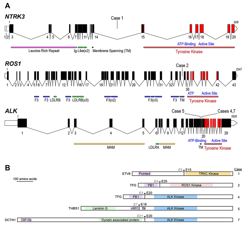
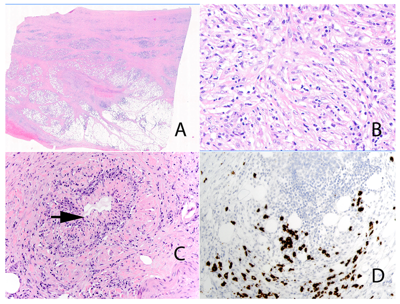
Inflammatory myofibroblastic tumor (IMT), a locally aggressive neoplasm capable of metastasis, may show an immunoglobulin (Ig)G4-rich lymphoplasmacytic infiltrate. Prior reports suggest that storiform-fibrosis and obliterative phlebitis aid in the distinction of IMT from IgG4-related diseases. Herein, we highlight the morphologic overlap between the 2 diseases, and emphasize the importance of a multiplex fusion assay in the distinction of IgG4-related disease (IgG4-RD) from IMT. We identified 7 IMTs with morphologic and immunohistochemical features of IgG4-RD; 3 patients were originally diagnosed with IgG4-RD. Demographic, clinical and morphologic data was recorded. We also reevaluated 56 patients with IgG4-RD. We performed immunohistochemistry for IgG4, IgG, ALK, and ROS1. In situ hybridization for IgG4 and IgG was performed in selected cases. A multiplex next-generation sequencing-based RNA assay for gene fusions was performed to detect all known IMT-related gene fusions. All 7 IMTs showed a dense lymphoplasmacytic infiltrate and storiform-type fibrosis, with obliterative phlebitis noted in 3 cases. The neoplastic stromal cells constituted <5% of overall cellularity and stromal atypia was either absent or focal and mild. Elevated numbers of IgG4 positive cells and increased IgG4 to IgG ratio was identified in all cases. Four cases showed ALK related abnormalities: 3 fusions and one alternative transcription initiation; while 2 patients showed ROS1 and NTRK3 fusions. One tumor was negative for known IMT-related gene fusions. All 56 IgG4-RD cases were negative for ALK and ROS1 on immunohistochemistry; 6 cases were negative on the fusion assay. Highly inflamed IMTs are indistinguishable from IgG4-RD both histologically and on immunohistochemistry for IgG4. We advocate scrutinizing patients with presumptive single organ IgG4-RD for IMT and the diagnostic algorithm should include ALK and ROS1 immunohistochemistry and, in selected cases, a next-generation sequencing-based fusion assay that covers known IMT-associated gene fusions.


Figures

Figure 1:
Figure 1:
Inflammatory myofibroblastic tumor with TFG-ROS1 fusion (case 2). Biopsy on Panel A and B, thoracoscopic biopsy on Panel C, D, E and F. The needle biopsy (Panel A and B) showed a dense plasma cell infiltrate and elevated numbers of IgG4 positive cells (not illustrated). The thoracoscopic biopsy (panel C and inset) although less inflamed, shows prominent stromal cells with atypia. Diffuse increase in IgG4 positive plasma cells (immunohistochemical stain for IgG4) (Panel D). Panel E inset shows a focus of obliterative phlebitis (* highlights the outlines of the obliterated vein). The obliterated vein is highlighted on an elastic stain (Panel F).
Figure 2.
Figure 2.
Inflammatory myofibroblastic tumor (case 5) with THBS1-ALK fusion. Storiform-type fibrosis (Panel A). * marks the center with fascicles of fibroinflammatory cells (arrow) emanating from this region. The neoplastic cells are diffusely positive for ALK (Panel B) Note the occasional stromal cells with vesicular nuclei (Panel B, inset). The plasma cells are diffusely positive for IgG4 in-situ hybridization (Panel C and inset) and IgG in-situ hybridization (Panel D and inset). The IgG4 to IgG ratio was > 90%.
Figure 3.
Figure 3.
Inflammatory myofibroblastic tumor with ETV6-NTRK3 fusion (case 1). The lung lesion is well circumscribed (Panel A). Note the storiform-type fibrosis (Panel B). On high-power the inflammatory cells dominate with only occasional stromal cells with vesicular nuclei (Panel C); storiform-type fibrosis is also seen. Immunohistochemical stain for IgG4 showing an IgG4-rich infiltrate (Panel D).
Figure 4:
Figure 4:
Gastric inflammatory myofibroblastic tumor (case #6), Panel A-D. The tumor involved the submucosa and muscularis propria (Panel A) and showed a prominent storiform-type pattern (Panel B). Occasional larger cells with vesicular nuclei are present (Panel C). These cells are diffusely positive for ALK (Panel D, immunohistochemistry). Tracheal tumor, likely representing an inflammatory myofibroblastic tumor (case #3) (Panel E and F). The tumor shows a prominent storiform-type pattern (Panel E). Note the stromal cells, similar to those depicted in figures 1–3 (Panel F) (arrow). However, the fusion panel was negative for IMT-related genetic alterations.
Figure 5:
Figure 5:
A Schematic of the human ALK, ROS1, and NTRK3 loci. Exon numbers are shown below their respective boxes for RefSeq ALK transcript variant 1 (NM_004304.4), ROS1 (NM_006180.4), and NTRK3 transcript variant 1 (NM_001012338.2). Fusion breakpoints are shown as dotted lines for the indicated cases; all breakpoints are intronic. Note that the three schematics are not at the same scale (amino acid residue numbers are listed above first and last exons), exons are drawn at a larger scale than introns, and introns are not drawn to the same scale for each gene (ALK locus is ~739 kB, ROS1 is ~137 kB, and NTRK3 is ~384 kB). Domains LDLR: LDL Receptor (A/B); F3: Fibronectin type III; MAM: meprin, A-5, mu-receptor; TM: Transmembrane Helix). B: Schematic of predicted fusion protein products (see also Table 1). Triangles and Ex notation indicate the fusion breakpoints, preceding partner gene exon, and subsequent kinase exon. Purple shaded domains are those predicted or shown to induce dimerization or trimerization in the fusion partner (Pointed: sterile alpha motif (SAM) / helix loop helix (HLH) oligomerization domain; PB1: Phox and Bem1p interaction domain; vWFC von Willebrand Factor C domain; CAP-Gly: cytoskeleton-associated protein glycine-rich domain); Green shaded domains are other annotated sequence features. Gray shaded TM: transmembrane domain. Proteins are drawn to scale (DCTN1-ALK fusion = 1383 AA).
Figure 6:
Figure 6:
IgG4-related retroperitoneal fibrosis. Low power view (Panel A). High-power view highlights the prominent population of stromal cells with mild nuclear atypia in the background of lymphocytes and plasma cells (Panel B). Obliterative phlebitis (Panel C) (arrow). Immunohistochemical stain for IgG4 showing increased numbers of IgG4 positive plasma cells (Panel D).

References

    1. Antonescu CR, Suurmeijer AJ, Zhang L, et al. Molecular characterization of inflammatory myofibroblastic tumors with frequent ALK and ROS1 gene fusions and rare novel RET rearrangement. Am J Surg Pathol. 2015;39:957–967. - PMC - PubMed
    1. Hornick JL, Sholl LM, Dal Cin P, et al. Expression of ROS1 predicts ROS1 gene rearrangement in inflammatory myofibroblastic tumors. Mod Pathol. 2015;28:732–739. - PMC - PubMed
    1. Lovly CM, Gupta A, Lipson D, et al. Inflammatory myofibroblastic tumors harbor multiple potentially actionable kinase fusions. Cancer Discov. 2014;4:889–895. - PMC - PubMed
    1. Yamamoto H, Yoshida A, Taguchi K, et al. ALK, ROS1 and NTRK3 gene rearrangements in inflammatory myofibroblastic tumours. Histopathology. 2016;69:72–83. - PubMed
    1. Gleason BC, Hornick JL. Inflammatory myofibroblastic tumours: where are we now? J Clin Pathol. 2008;61:428–437. - PubMed

Substances