Scotoma Simulation in Healthy Subjects
- PMID: 30451808
- PMCID: PMC6266299
- DOI: 10.1097/OPX.0000000000001310
Scotoma Simulation in Healthy Subjects
Abstract
Significance: This article shows a successful concept for simulating central scotoma, which is associated with age-related macular degeneration (AMD), in healthy subjects by an induced dark spot at the retina using occlusive contact lenses. The new concept includes a control mechanism to adjust the scotoma size through controlling pupil size without medication. Therefore, a miniaturized full-field adaptation device was used.
Purpose: The aim of this study was to design a novel concept to simulate AMD scotoma in healthy subjects using occlusive contact lenses.
Methods: To define an optimal set of lens parameters, we constructed an optical model and considered both the anatomical pupil diameter and the opaque central zone diameter of the contact lens. To adjust the scotoma size, we built a miniaturized full-field adaptation device. We demonstrate the validity of this novel concept by functional measurements of visual fields using automated threshold perimetry. Finally, we conducted a perception study including two tasks, consisting of pictograms and letters. The stimuli were presented at different eccentricities and magnifications.
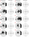
Results: The visual fields of all 10 volunteers exhibited absolute scotomas. The loss of contrast sensitivity ranged within 27 and 36 dB (P < .05), and the scotoma localizations were nearly centered to the macula (mean variation, 2.0 ± 4.8° horizontally; 3.5 ± 4.7° vertically). The eccentric perception of letters showed the largest numbers of correctly identified stimuli. The perception of pictograms showed significantly reduced numbers (P < .0001) and revealed a dependency on magnification. The results suggest that best perception is possible for magnified stimuli near the scotoma.
Conclusions: We demonstrated that the creation of an absolute simulated AMD scotoma is possible using occlusive contact lenses combined with a miniaturized full-field adaptation device.
Conflict of interest statement
Figures


Similar articles
-
Simulation of a central scotoma using contact lenses with an opaque centre.Ophthalmic Physiol Opt. 2018 Jan;38(1):76-87. doi: 10.1111/opo.12422. Ophthalmic Physiol Opt. 2018. PMID: 29265475 Free PMC article.
-
Simulation contact lenses for AMD health state utility values in NICE appraisals: a different reality.Br J Ophthalmol. 2015 Apr;99(4):540-4. doi: 10.1136/bjophthalmol-2014-305802. Epub 2014 Oct 28. Br J Ophthalmol. 2015. PMID: 25351679 Free PMC article.
-
Rehabilitative approach in patients with ring scotoma.Can J Ophthalmol. 2013 Oct;48(5):420-6. doi: 10.1016/j.jcjo.2013.07.012. Can J Ophthalmol. 2013. PMID: 24093190
-
Visual function in patients with choroidal neovascularization resulting from age-related macular degeneration: the importance of looking beyond visual acuity.Optom Vis Sci. 2006 Mar;83(3):178-89. doi: 10.1097/01.opx.0000204510.08026.7f. Optom Vis Sci. 2006. PMID: 16534460 Review.
-
Functional and cortical adaptations to central vision loss.Vis Neurosci. 2005 Mar-Apr;22(2):187-201. doi: 10.1017/S0952523805222071. Vis Neurosci. 2005. PMID: 15935111 Free PMC article. Review.
Cited by
-
Customizing spatial remapping of letters to aid reading in the presence of a simulated central field loss.J Vis. 2024 Apr 1;24(4):17. doi: 10.1167/jov.24.4.17. J Vis. 2024. PMID: 38635281 Free PMC article.
-
Opportunities and Limitations of a Gaze-Contingent Display to Simulate Visual Field Loss in Driving Simulator Studies.Front Neuroergon. 2022 Jun 10;3:916169. doi: 10.3389/fnrgo.2022.916169. eCollection 2022. Front Neuroergon. 2022. PMID: 38235462 Free PMC article.
-
Simulating Macular Degeneration to Investigate Activities of Daily Living: A Systematic Review.Front Neurosci. 2021 Aug 13;15:663062. doi: 10.3389/fnins.2021.663062. eCollection 2021. Front Neurosci. 2021. PMID: 34483815 Free PMC article.
References
-
- Jonas JB, Cheung CMG, Panda-Jonas S. Updates on the Epidemiology of Age-related Macular Degeneration. Asia Pac J Ophthalmol (Phila) 2017;6:493–7. - PubMed
-
- Jonas JB, Bourne RR, White RA, et al. Visual Impairment and Blindness Due to Macular Diseases Globally: A Systematic Review and Meta-analysis. Am J Ophthalmol 2014;158:808–15. - PubMed
Publication types
MeSH terms
LinkOut - more resources
Full Text Sources
Medical
Miscellaneous

