. 2018 Nov 19;9(1):4881.
doi: 10.1038/s41467-018-07335-2.

Hidden diversity of soil giant viruses

Affiliations

Hidden diversity of soil giant viruses

Frederik Schulz et al. Nat Commun. .

Abstract

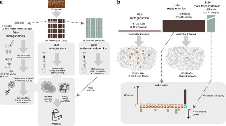
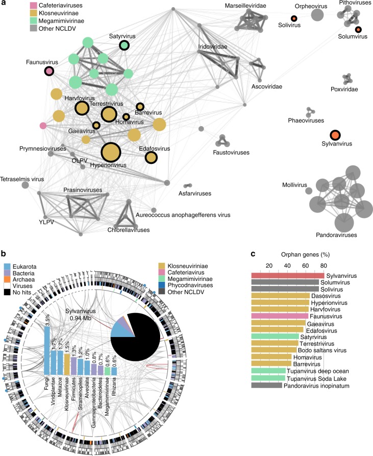
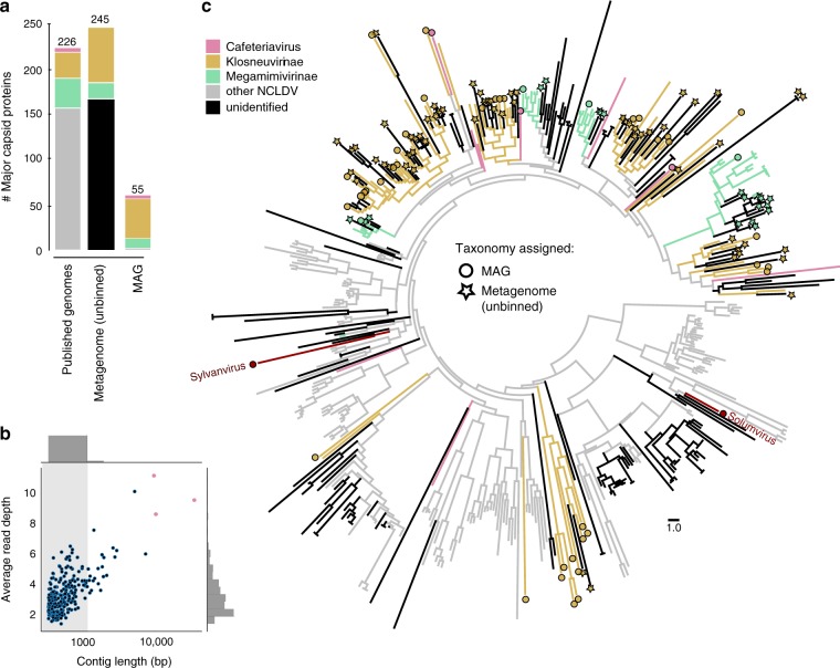
Known giant virus diversity is currently skewed towards viruses isolated from aquatic environments and cultivated in the laboratory. Here, we employ cultivation-independent metagenomics and mini-metagenomics on soils from the Harvard Forest, leading to the discovery of 16 novel giant viruses, chiefly recovered by mini-metagenomics. The candidate viruses greatly expand phylogenetic diversity of known giant viruses and either represented novel lineages or are affiliated with klosneuviruses, Cafeteria roenbergensis virus or tupanviruses. One assembled genome with a size of 2.4 Mb represents the largest currently known viral genome in the Mimiviridae, and others encode up to 80% orphan genes. In addition, we find more than 240 major capsid proteins encoded on unbinned metagenome fragments, further indicating that giant viruses are underexplored in soil ecosystems. The fact that most of these novel viruses evaded detection in bulk metagenomes suggests that mini-metagenomics could be a valuable approach to unearth viral giants.


Conflict of interest statement

The authors declare no competing interests.

Figures

Fig. 1
Fig. 1
Discovery pipeline for soil giant viruses. a Overall workflow. Fourteen forest soil cores from Barre Woods long-term experimental warming site were sub-sampled into organic horizon and mineral zone resulting in 28 total samples. Total DNA and RNA were extracted from 28 soil samples for bulk metagenomics and metatranscriptomics. Of these samples, a subset of four encompassing two organic and two mineral layers were selected for flow-sorted mini-metagenomics. Cells and presumably viral particles, were separated from soil, stained with SYBR green nucleic acid stain and sorted using fluorescence activated cell sorting (FACS). Ninety sorted pools of 100 SYBR+ particles underwent lysis, whole genome amplification, library preparation, and sequencing on the Illumina NextSeq platform. Phylogenomic analysis of metagenome assembled genomes (MAGs) facilitated the identification of novel giant viruses. There was no correlation of presence or absence of giant viruses and sample treatment (Supplementary Table 3). b Data analysis summary. Fifteen giant virus MAGs (orange circles) were recovered from sorted samples, while only one giant virus MAG (turquoise circle) was recovered from the bulk metagenomes. The other 1778 MAGs from the mini-metagenomes (gray circles) and 1772 MAGs from the bulk metagenomes (gray circles) were of bacterial or archaeal origin and not analyzed further in this study. Mapping of bulk metagenome reads to MAGs revealed ~9× coverage of the bulk-metagenome derived MAG and <1× coverage of MAGs derived from mini-metagenomes, confirming the inability to recover these novel giant virus genomes using bulk metagenomics despite deep sequencing efforts. Assembly and mapping of metatranscriptome data indicated expression of only few of the novel giant virus genes of MAGs derived from mini-metagenomes
Fig. 2
Fig. 2
Expansion of NCLDV diversity by novel soil giant viruses. a Phylogenetic tree (IQ-tree LG+F+R6) of NCLDV inferred from a concatenated protein alignment of five core nucleocytoplasmic virus orthologous genes (NCVOGs). The tree was built from a representative set of NCDLV genomes after de-replication by ANI clustering (95% id). Novel soil NCLDV lineages and existing major NCLDV lineages grouping together with soil NCLDV are highlighted in black. The scale bar represents substitutions per site. Branch support values are shown in data S1. Branches are collapsed if support was low (<50), filled circles indicate moderate support (50–80, white) or high support (80–97, black), branches without circles are fully supported (>97). b Detailed phylogenetic tree of the Mimiviridae. Diameter of filled circles correlates with assembly size and shades of gray with GC% ranging from 20% (light gray) to 60% (dark gray). Bar plots summarize total number of encoded aminoacyl-tRNA synthetases (aaRS) and tRNAs. In addition, completeness was estimated based on number of identified marker genes out of 20 ancestral NCVOGs (more details are shown in Supplementary Fig. 1). c Increase of phylogenetic diversity (PD) after adding the soil NCLDV MAGs (black) to representative sets of NCLDV reference genomes (gray). Naming considerations are shown in Supplementary Table 2
Fig. 3
Fig. 3
Genome novelty of soil giant viruses. a Nucleocytoplasmic large DNA virus (NCLDV) gene sharing network, with nodes representing genomes, node diameter correlating with genome size, edge diameter and color intensity with normalized percentage of genes in shared gene families between node pairs above a threshold of 18%. b Circular representation of the sylvanvirus genome. From outside to inside: Blue filled circles depict location of encoded tRNAs. The second ring displays positions of genes (gray) either on the minus or the plus strand. The next track illustrates GC content in shades of gray ranging from 20% (white) to 60% (dark gray). The fourth track shows color-coded origin of proteins with best blastp hits (e-value 1e−5) to cellular homologs. Best hits against viral proteins are indicated in white and if possible, further broken down based on their taxonomic origin color-coded on the most inner track. Finally, lines in the middle of the plot connect paralogs (gray) and nearly identical repeats (orange). The pi chart in the center of the plot summarizes the percentage of genes with and without cellular homologs, which are further broken down based on best blastp hits (e-value 1e−5) hits in the adjacent bar plot. c Percentage of genes in NCLDV genomes with bacterial or eukaryotic homologs and with no blastp hits (e-value 1e−5) in the NCBI nr database, highlighting the unique position of sylvanvirus
Fig. 4
Fig. 4
Hidden diversity of giant viruses in bulk metagenomes. a Total number of major capsid proteins (MCPs) found in reference nucleocytoplasmic large DNA virus (NCLDV) genomes, metagenome assembled genomes (MAGs), or recovered from bulk metagenomes on contigs >1 kb and contigs <1 kb (dark gray), colored by taxonomy. b Size and cover of bulk metagenome contigs containing MCP genes, either from the unbinned fraction (filled blue circles) or the MAGs (filled pink circles). c Phylogenetic tree of the MCPs of NCLDV. Branches are color-coded based on taxonomic origin of MCPs inferred by relationship in the tree to MCPs of known reference NCLDV. MCPs of novel giant viruses from this study which are not members of the Mimiviridae are indicated in red. Branches labeled with a circle represent novel MCP from MAGs generated in this study while stars indicate MCPs recovered from the unassembled fraction (contigs >1 kb) of bulk-metagenomes. Circles and stars are filled in color if taxonomy could be assigned based on the tree and in black if it was not possible to assign taxonomy

References

    1. Aherfi S, Colson P, La Scola B, Raoult D. Giant viruses of amoebas: an update. Front. Microbiol. 2016;7:349. doi: 10.3389/fmicb.2016.00349. - DOI - PMC - PubMed
    1. Andrade ACDSP, et al. Ubiquitous giants: a plethora of giant viruses found in Brazil and Antarctica. Virol. J. 2018;15:22. doi: 10.1186/s12985-018-0930-x. - DOI - PMC - PubMed
    1. Pagnier I, et al. A decade of improvements in Mimiviridae and Marseilleviridae isolation from amoeba. Intervirology. 2013;56:354–363. doi: 10.1159/000354556. - DOI - PubMed
    1. Yoosuf N, et al. Draft genome sequences of Terra1 and Terra2 viruses, new members of the family Mimiviridae isolated from soil. Virology. 2014;452-453:125–132. doi: 10.1016/j.virol.2013.12.032. - DOI - PubMed
    1. Boughalmi M, et al. High-throughput isolation of giant viruses of the Mimiviridae and Marseilleviridae families in the Tunisian environment. Environ. Microbiol. 2013;15:2000–2007. doi: 10.1111/1462-2920.12068. - DOI - PubMed

Publication types