Review
. 2019 Jan;15(1):40-52.
doi: 10.1038/s41582-018-0101-0.

POLG-related disorders and their neurological manifestations

Affiliations
Review

POLG-related disorders and their neurological manifestations

Shamima Rahman et al. Nat Rev Neurol. 2019 Jan.

Abstract

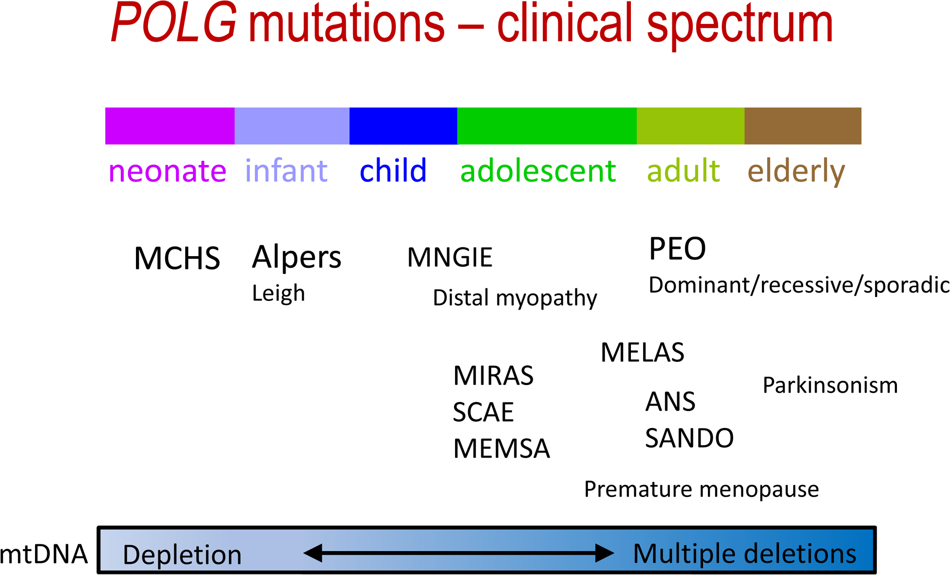
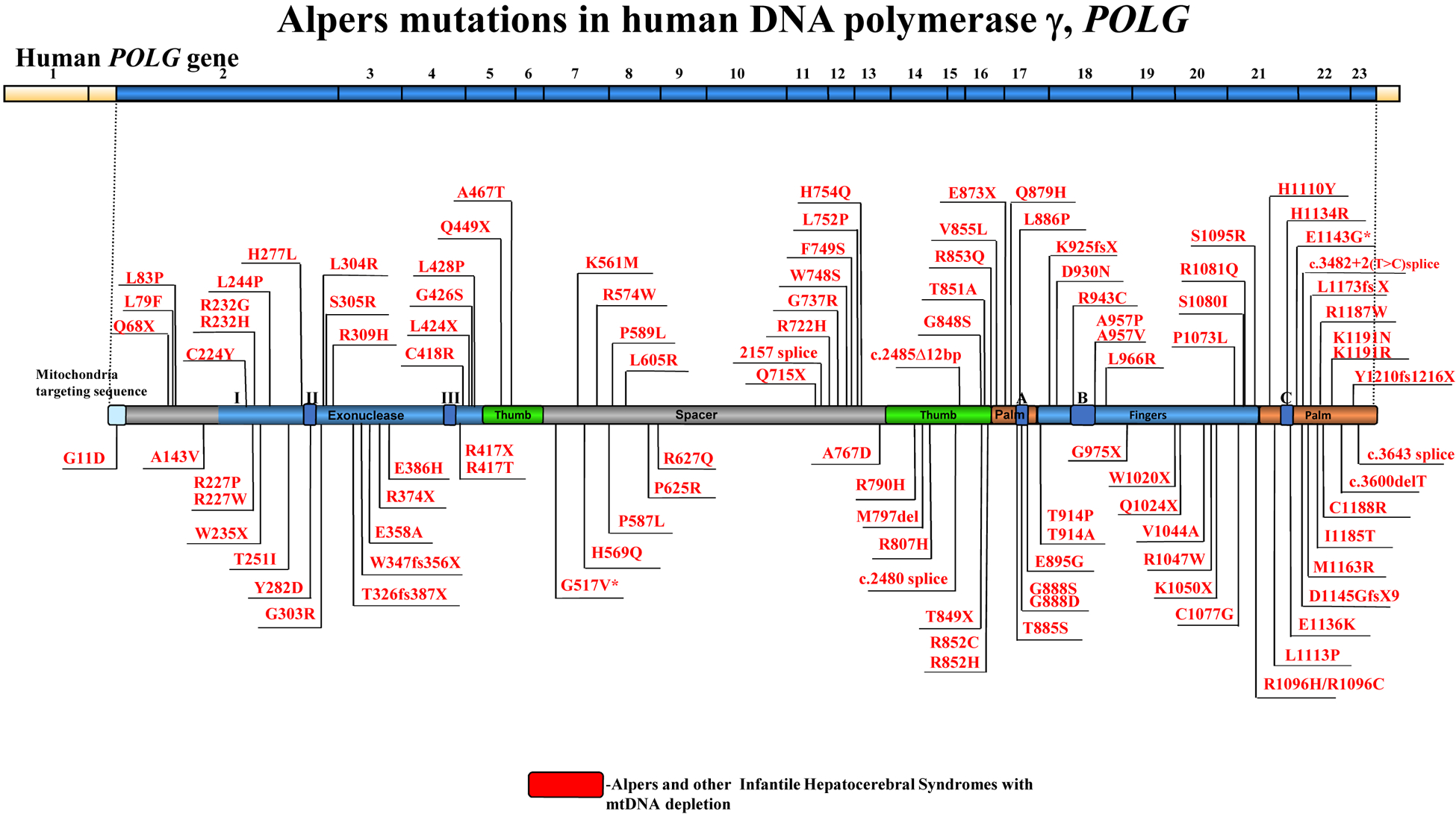
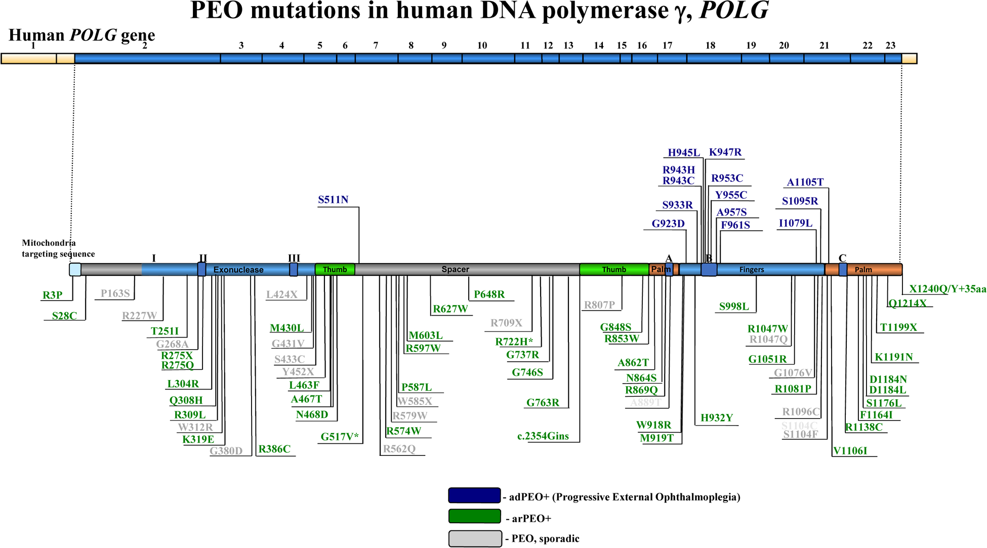
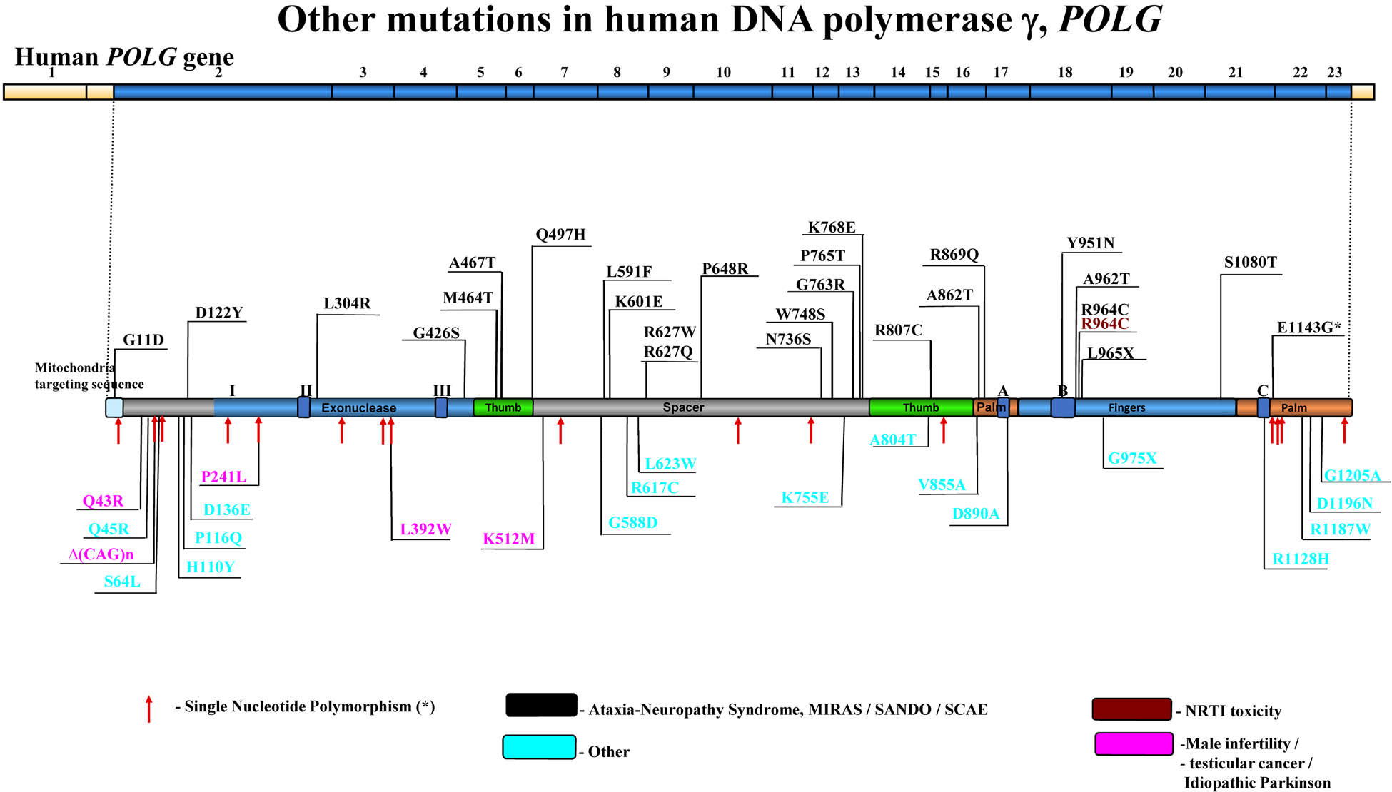
The POLG gene encodes the mitochondrial DNA polymerase that is responsible for replication of the mitochondrial genome. Mutations in POLG can cause early childhood mitochondrial DNA (mtDNA) depletion syndromes or later-onset syndromes arising from mtDNA deletions. POLG mutations are the most common cause of inherited mitochondrial disorders, with as many as 2% of the population carrying these mutations. POLG-related disorders comprise a continuum of overlapping phenotypes with onset from infancy to late adulthood. The six leading disorders caused by POLG mutations are Alpers-Huttenlocher syndrome, which is one of the most severe phenotypes; childhood myocerebrohepatopathy spectrum, which presents within the first 3 years of life; myoclonic epilepsy myopathy sensory ataxia; ataxia neuropathy spectrum; autosomal recessive progressive external ophthalmoplegia; and autosomal dominant progressive external ophthalmoplegia. This Review describes the clinical features, pathophysiology, natural history and treatment of POLG-related disorders, focusing particularly on the neurological manifestations of these conditions.


Conflict of interest statement

Competing interests

The authors declare no competing interests.

Figures

Figure 1 |
Figure 1 |
The clinical spectrum of POLG-related disease. Clinical spectrum of POLG-related disease according to age of onset, and the defects (mitochondrial DNA (mtDNA) depletion or deletions) associated with the diseases. AHS, Alpers–Huttenlocher syndrome; ANS, ataxia neuropathy spectrum; MCHS, myocerebrohepatopathy spectrum; MELAS, mitochondrial encephalomyopathy, lactic acidosis and stroke-like episodes; MEMSA, myoclonic epilepsy myopathy sensory ataxia; MNGIE, mitochondrial neurogastrointestinal encephalopathy; PEO, progressive external ophthalmoplegia; SANDO, sensory ataxia neuropathy dysarthria and ophthalmoplegia; SCAE, spinocerebellar ataxia with epilepsy.
Figure 2 |
Figure 2 |
POLG mutations. Mutation map depicting disease-associated amino acid substitutions on the primary structure of POLG. In each panel, the top line depicts the 23 exons of the cDNA and the lower line represents the linear polypeptide with the functional domains (exonuclease and polymerase) indicated. The polymerase active site is subdivided into thumb, palm and fingers subdomains. A full list of disease-related substitutions can be found in the Human DNA Polymerase Gamma Mutation Database. Asterisks indicate mutations that have also been identified as frequent single nucleotide polymorphisms.a | Mutations associated with Alpers–Huttenlocher syndrome and other infantile hepatocerebral syndromes that cause mitochondrial DNA depletion. b | Mutations associated with progressive external ophthalmoplegia (PEO). c | Other mutations linked to POLG-related disease. ANS, ataxia neuropathy spectrum; MIRAS, mitochondrial recessive ataxia syndrome; NRTI, nucleoside reverse transcriptase inhibitor; SANDO, sensory ataxia neuropathy dysarthria and ophthalmoplegia; SCAE, spinocerebellar ataxia with epilepsy.
Figure 2 |
Figure 2 |
POLG mutations. Mutation map depicting disease-associated amino acid substitutions on the primary structure of POLG. In each panel, the top line depicts the 23 exons of the cDNA and the lower line represents the linear polypeptide with the functional domains (exonuclease and polymerase) indicated. The polymerase active site is subdivided into thumb, palm and fingers subdomains. A full list of disease-related substitutions can be found in the Human DNA Polymerase Gamma Mutation Database. Asterisks indicate mutations that have also been identified as frequent single nucleotide polymorphisms.a | Mutations associated with Alpers–Huttenlocher syndrome and other infantile hepatocerebral syndromes that cause mitochondrial DNA depletion. b | Mutations associated with progressive external ophthalmoplegia (PEO). c | Other mutations linked to POLG-related disease. ANS, ataxia neuropathy spectrum; MIRAS, mitochondrial recessive ataxia syndrome; NRTI, nucleoside reverse transcriptase inhibitor; SANDO, sensory ataxia neuropathy dysarthria and ophthalmoplegia; SCAE, spinocerebellar ataxia with epilepsy.

References

    1. Kukat C et al. Super-resolution microscopy reveals that mammalian mitochondrial nucleoids have a uniform size and frequently contain a single copy of mtDNA. Proc Natl Acad Sci USA 108 13534–13539, (2011). - PMC - PubMed
    1. Korhonen JA, Pham XH, Pellegrini M & Falkenberg M Reconstitution of a minimal mtDNA replisome in vitro. EMBO J 23, 2423–2429 (2004). - PMC - PubMed
    1. Copeland WC & Longley MJ Mitochondrial genome maintenance in health and disease. DNA Repair (Amst) 19, 190–198 (2014). - PMC - PubMed
    1. Bebenek K & Kunkel TA Functions of DNA polymerases. Adv Protein Chem 69, 137–165 (2004). - PubMed
    1. Ropp PA & Copeland WC Cloning and characterization of the human mitochondrial DNA polymerase, DNA polymerase gamma. Genomics 36, 449–458 (1996). - PubMed

Publication types

MeSH terms