. 2018 Dec 18;57(50):6878-6887.
doi: 10.1021/acs.biochem.8b01032. Epub 2018 Dec 4.

RNA Recognition-like Motifs Activate a Mitogen-Activated Protein Kinase

Affiliations

RNA Recognition-like Motifs Activate a Mitogen-Activated Protein Kinase

Timothy Phillips et al. Biochemistry. .

Abstract

Smk1 is a mitogen-activated protein kinase (MAPK) family member in the yeast Saccharomyces cerevisiae that controls the postmeiotic program of spore formation. Ssp2 is a meiosis-specific protein that activates Smk1 and triggers the autophosphorylation of its activation loop. A fragment of Ssp2 that is sufficient to activate Smk1 contains two segments that resemble RNA recognition motifs (RRMs). Mutations in either of these motifs eliminated Ssp2's ability to activate Smk1. In contrast, deletions and insertions within the segment linking the RRM-like motifs only partially reduced the activity of Ssp2. Moreover, when the two RRM-like motifs were expressed as separate proteins in bacteria, they activated Smk1. We also find that both motifs can be cross-linked to Smk1 and that at least one of the motifs binds near the ATP-binding pocket of the MAPK. These findings demonstrate that motifs related to RRMs can directly activate protein kinases.


Conflict of interest statement

The authors declare no competing financial interest.

Figures

Figure 1.
Figure 1.
(A) Diagram of Ssp2 showing the TD and the KAD (top). The KAD is further subdivided into KARL1 (turquoise), the linker (yellow), and KARL2 (magenta). The KAD modeled after the structure of the polypyrimidine tract-binding protein (middle) and a secondary structure model of the KAD predicted by PSIPRED (below) are shown with Ssp2 residue numbers in the linker indicated. The positions of F211 and F327, corresponding to the F residues in canonical RRMs that stack with RNA bases, and F307, a residue that is present in a subset of RRMs, are indicated with upward arrows. (B) Mutational analyses of F211, F307, and F327 in Ssp2. Bacteria were transformed with plasmids that direct the expression of Smk1 and either wild-type or mutant forms of Ssp2KAD-GST as indicated. The most leftward lane shows bacterial cells analyzed in the absence of an inducer (–) as a control. Transformants were induced and total cellular extracts analyzed by immunoblots using the indicated antibodies. The lanes correspond to the following plasmids: WT, pJT118; Δ, pJT114; F307A/F327A, pAR10; F307Y/F327Y, pAR11; F307A, pAR4; F307Y, pAR6; F327A, pAR7; F327Y, pAR9; F211A, pAR1; F211Y, pAR3 (Table 2).
Figure 2.
Figure 2.
KARL1 and KARL2 are functional when expressed as separate proteins. Bacteria producing residues 162–265 of Ssp2 fused to the C-terminus of MBP (KARL1), residues 266–371 of Ssp2 fused to the N-terminus of GST (KARL2), Ssp2KAD-GST (KAD), and Smk1, as indicated above, were assayed by immunoblotting using the antisera against the proteins indicated on the right. The KARL2 deletion mutants diagrammed below (vertical lines indicating deletion end points) were co-expressed with the functional MBP-Ssp2162–265 KARL1 protein, and the KARL1 deletions diagrammed below were co-expressed with the functional Ssp2266–371-GST (KARL2) protein. The lanes correspond to the following plasmids: lane 1, pTP39 and pTP36; lane 2, pJT114 and pACYDuet-1 (empty vector); lane 3, pJT118 and pACYDuet-1; lane 4, pTP35 and pTP14; lane 5, pTP35 and pTP41; lane 6, pTP35 and pTP15; lane 7, pTP39 and pTP14; lane 8, pTP40 and pTP14; lane 9, pTP44 and pTP14; lane 10, pTP45 and pTP14 (Table 2).
Figure 3.
Figure 3.
The SSP2 linker can tolerate substantial mutation. (A) Diagram of linker mutants L1–L4 in Ssp2. (B) Diploid SMK1-HH/SMK1-HH yeast containing one chromosomal copy of the indicated SSP2KAD-GST linker derivatives were transferred to sporulation medium, and cells were collected 8.5 h post-induction (when >75% of cells had completed MII). Smk1-HH was purified using nickel beads (NTA), and the fraction of Smk1-HH that had autophosphorylated Y209 was scored using pY209 and HA (Smk1) antibodies as indicated. The Ssp2-GST derivatives were purified from a portion of the extracts using glutathione agarose (GSH), and the relative fraction of Smk1 that was bound to Ssp2 was scored using antibodies against HA (Smk1) and GST (Ssp2). The input serves as a control for the amount of Smk1 and Ssp2 in the extracts. (C) The fraction of cells that completed the sporulation program was scored by counting asci that contained two or more refractile spores (dark bars) and are plotted with the fraction of Smk1 that had autophosphorylated Y209 (relative pY209:Smk1 ratios with the wild-type SSP2KAD-GST strain taken to be 1.0) (lighter gray bars). The following yeast strains were used in these experiments: JTY88 (WT), TPY13 (ssp2Δ), TPY29 (ssp2-L1), TPY28 (ssp2-L2), TPY30 (ssp2-L3), and TPY36 (ssp2-L4) (Table 1).
Figure 4.
Figure 4.
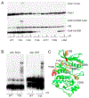
Residue near the ATP-binding pocket of Smk1 that is within 4 Å of Ssp2. (A) Smk1 derivatives containing the cross-linkable amino acid, Bpa, at the indicated positions were co-expressed in bacteria with Ssp2KAD-GST. Complexes were purified using reduced glutathione, and one half of the preparation was irradiated with UV light (+) and the other half was untreated (−). Samples were analyzed by electrophoresis through denaturing electrophoretic gels and immunoblotting using a Smk1 antibody (top) or a pY209 antiserum (bottom). The cross-linked (X-link) species migrates at approximately 130 kDa, a molecular weight slightly larger than the predicted molecular weight of a Smk1/Ssp2KAD-GST complex. NS (nonspecific) indicates a bacterial protein that cross-reacts with the pY209 antiserum. (B) Samples were processed as described for panel A and assayed using a GST or a Smk1 antiserum as indicated. All of the bacterial strains in these experiments contained pEVOL (provides the tRNA/synthetase pair that permits incorporation of Bpa at amber suppressible codons) and pJT115 (WT) or the derivatives of pJT115-containing amber codons at the indicated positions as listed in Table 2. (C) Smk1 modeled after ERK5. Positions substituted with Bpa and analyzed in panels A and B are indicated (tan). Residues of Smk1 that correspond to residues in Fus3 that interact with a peptide from Ste5 that triggers autophosphorylation of Fus3’s activation-loop Y are colored red (see Discussion).
Figure 5.
Figure 5.
Residues in both KARL1 and KARL2 are within 4 Å of Smk1. Ssp2KAD-GST derivatives containing Bpa at the indicated positions in (A) KARL1 or (B) KARL2 were co-expressed with Smk1 in bacteria. Complexes were purified using reduced glutathione beads and exposed to UV light (+) or left untreated (−). Samples were then analyzed by denaturing electrophoresis and immunobloting using Smk1 and Smk1-Y209p antisera. All of the bacterial strains in these experiments contained pEVOL (provides the tRNA/synthetase pair that permits incorporation of Bpa at amber suppressible codons) and pJT115 (WT) or the derivatives of pJT115-containing amber codons at the indicated positions as listed in Table 2. The Smk1 Y38 samples, which contain Bpa at residue 38 of Smk1, and the wild-type (WT) sample lacking Bpa are controls for cross-linking.

Similar articles

Cited by

References

    1. Chen RE, and Thorner J (2007) Function and regulation in MAPK signaling pathways: lessons learned from the yeast Saccharomyces cerevisiae. Biochim. Biophys. Acta, Mol. Cell Res 1773, 1311–1340. - PMC - PubMed
    1. Coulombe P, and Meloche S (2007) Atypical mitogen-activated protein kinases: structure, regulation and functions. Biochim. Biophys. Acta, Mol. Cell Res 1773, 1376–1387. - PubMed
    1. Pimienta G, and Pascual J (2007) Canonical and alternative MAPK signaling. Cell Cycle 6, 2628–2632. - PubMed
    1. Salvador JM, Mittelstadt PR, Guszczynski T, Copeland TD, Yamaguchi H, Appella E, Fornace AJ Jr., and Ashwell JD (2005) Alternative p38 activation pathway mediated by T cell receptor-proximal tyrosine kinases. Nat. Immunol 6, 390–395. - PubMed
    1. Ge B, Gram H, Di Padova F, Huang B, New L, Ulevitch RJ, Luo Y, and Han J (2002) MAPKK-independent activation of p38alpha mediated by TAB1-dependent autophosphorylation of p38alpha. Science 295, 1291–1294. - PubMed

Publication types

MeSH terms

Substances