. 2018 Nov 19;13(11):e0207417.
doi: 10.1371/journal.pone.0207417. eCollection 2018.

Selective inhibition of carbonic anhydrase IX over carbonic anhydrase XII in breast cancer cells using benzene sulfonamides: Disconnect between activity and growth inhibition

Affiliations

Selective inhibition of carbonic anhydrase IX over carbonic anhydrase XII in breast cancer cells using benzene sulfonamides: Disconnect between activity and growth inhibition

Mam Y Mboge et al. PLoS One. .

Retraction in

Abstract

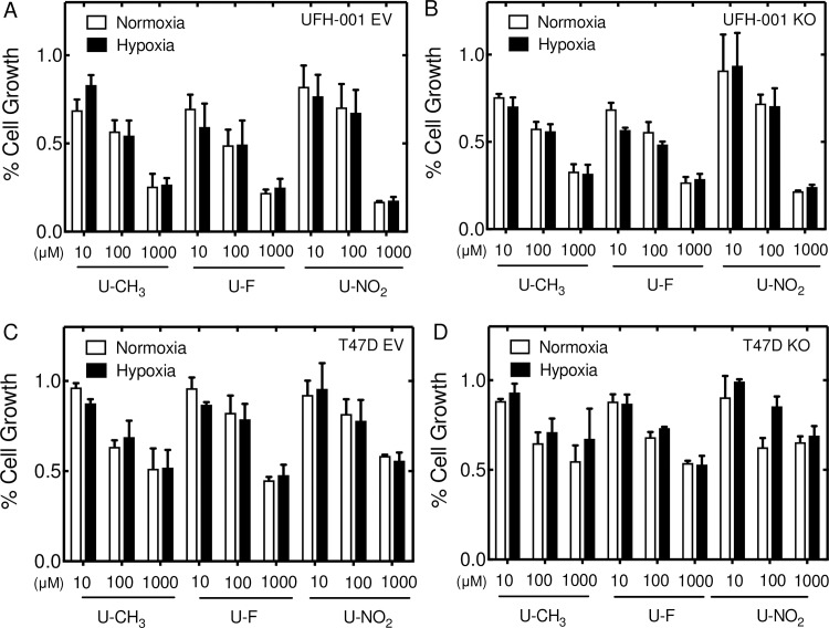
Carbonic anhydrases (CAs) have been linked to tumor progression, particularly membrane-bound CA isoform IX (CA IX). The role of CA IX in the context of breast cancer is to regulate the pH of the tumor microenvironment. In contrast to CA IX, expression of CA XII, specifically in breast cancer, is associated with better outcome despite performing the same catalytic function. In this study, we have structurally modeled the orientation of bound ureido-substituted benzene sulfonamides (USBs) within the active site of CA XII, in comparison to CA IX and cytosolic off-target CA II, to understand isoform specific inhibition. This has identified specific residues within the CA active site, which differ between isoforms that are important for inhibitor binding and isoform specificity. The ability of these sulfonamides to block CA IX activity in breast cancer cells is less effective than their ability to block activity of the recombinant protein (by one to two orders of magnitude depending on the inhibitor). The same is true for CA XII activity but now they are two to three orders of magnitude less effective. Thus, there is significantly greater specificity for CA IX activity over CA XII. While the inhibitors block cell growth, without inducing cell death, this again occurs at two orders of magnitude above the Ki values for inhibition of CA IX and CA XII activity in their respective cell types. Surprisingly, the USBs inhibited cell growth even in cells where CA IX and CA XII expression was ablated. Despite the potential for these sulfonamides as chemotherapeutic agents, these data suggest that we reconsider the role of CA activity on growth potentiation.


Conflict of interest statement

The authors have declared that no competing interests exist.

Figures

Fig 1
Fig 1. USBs bound in the active site of CAs.
Panel A. Ribbon diagram of monomeric CA isoform II (gray). Panel C. Ribbon diagram of the catalytic domains of dimeric CA isoform IX (cyan). Panel E. Ribbon diagram of the catalytic domains of dimeric CA isoform XII (wheat). Surface representation of compounds U-CH3 (pink), U-F (green) and U-NO2 (yellow) in complex with monomers of CA II (Panel B), the CA IX-mimic (Panel D), and modeled into the active site of CA XII (Panel F). Catalytic zinc (magenta sphere), hydrophilic (blue) and hydrophobic (orange) residues are as shown. Red double-headed arrows indicate isoform specificity relative to residue 131 (labeled in white). These arrows also show flexibility in tail conformations seen in CA II and CA IX but not in CA XII. Previously published Ki values of each compound bound to purified CA II, CA IX and CA XII are noted next to the inhibitor name [67, 68]. Figures were designed using PyMol.
Fig 2
Fig 2. CA expression and activity in breast cell lines.
Panel A. Protein expression in a normal immortalized basal type breast cell line (MCF 10A) and a triple negative breast cancer (TNBC) cell line (UFH-001), using western blot analysis (under normoxic (N) or hypoxic conditions (H) for 16 h, respectively). Panel B. Immunoblots for CAs II, IX and XII protein expression in MCF 10A and the luminal ER positive breast cancer cell line (T47D). Panels C and D. UFH-001 and T47D cells were grown for 3 days at which point they were exposed to normoxia or 16 h of hypoxia and the cells assayed for CA IX and XII activity, respectively, using the MIMS assay (see S3A and S3B Fig for a description of a pictorial representation of the MIMS assay). First order rate constants for CA activity in UFH-001 cells (Panel A) and T47D cells (Panel B), as described in the methods, are shown. These data represent three independent experiments and are reported as the mean ± SEM.
Fig 3
Fig 3. Effects of USBs on CA IX and CA XII activity in breast cancer cell lines.
Panels A-C. UFH-001 cells were grown for 4 days under normoxic conditions. CA IX activity (0.5 × 10⁶ cells/mL, unless otherwise indicated) was assayed using MIMS in the presence or absence of U-CH3 (Panel A), U-F (Panel B) or U-NO2 (Panel C). Panels D-F. T47D cells prepared similarly to UFH-001 cells, but cultured for 6 days, were also assayed for CA XII activity (0.5 × 10⁶ cells/mL) in the absence or presence of the same USB based inhibitors: U-CH3 (Panel D), U-F (Panel E) or U-NO2 (Panel F). Atom fractions of 18O in CO2 were collected continuously. For ease of illustration, data points at 25-s intervals are shown. Phase I in UFH-001 cells indicate CA II activity and Phase II indicate CA IX activity. In T47D cells, the progress curves are linear and representative of CA XII activity. Arrows indicate time point at which cells were added. Panel G. Ki values of each compound in UFH-001 and T47D cells, under normoxic or hypoxic (16 h) conditions, are shown. Data are representative of the average of triplicate experiments ± SEM.
Fig 4
Fig 4. Effect of CA IX and CA XII knockdown on CA activity.
CA IX knockout in UFH-001 cells was accomplished using Crispr/Cas9 technology. CA XII knockdown in T47D cells was performed using shRNAi lentiviral strategies. Cells were grown similarly to those described in Fig 3. Panel A. CA activity was measured in 1 x 10⁶ UFH-001 cells (EV controls, CA IX KO, or cells treated with the pegylated sulfonamide, N-3500). Panel B. CA activity was measured in 5 x 10⁵ T47D cells (EV controls, CA XII KO, or cells treated with N-3500. Data represent duplicate experiments.
Fig 5
Fig 5. Effects of USBs on breast cancer cell growth.
Breast cancer cell lines grown under normal culture conditions for 24 h were exposed to compounds for 48 h [U-CH3 (Panel A), U-F (Panel B), U-NO2 (Panel D)] or 96 h [U-CH3 (Panel D), U-F (Panel E), or U-NO2 (Panel F)]. MTT assay was performed at 48 h and 96 h, respectively. Data shown are an average of at least three independent experiments and are represented as the mean ± SEM.
Fig 6
Fig 6. Effects of USBs on cell growth in the presence or absence of hypoxia or in CA knockout cells.
One day after plating, breast cancer cell lines were exposed to normoxic or hypoxic conditions for 48 h in the presence of U-CH3, U-F, or U-NO2 at the given concentrations in UFH-001 empty vector control (EV) cells (Panel A), UFH-001 CA IX KO (UFH-001 KO) cells (Panel B), T47D EV cells (Panel C) and T47D CA XII KO (T47D KO) cells (Panel D). MTT assay was performed and shown as % cell growth. Data shown represent at least three independent experiments and are shown as the mean ± SEM.
Fig 7
Fig 7. Effects of USBs on breast cancer cell viability.
The cytotoxic effects of USBs were evaluated using the LDH release assay. MCF 10A (Panel A), UFH-001 (Panel B) and T47D (Panel C) cells were grown in 96-well plates and exposed to U-CH3, U-F or U-NO2 for 48 h, under normoxic conditions. LDH release was assayed after treatment, results were evaluated, and data analyzed using Prism. Data shown are representative of three independent experiments and as the mean ± SEM, *p < 0.05.
Fig 8
Fig 8. Effects of USBs on cell cycle transition in breast cancer cells.
Cell cycle analysis was performed in UFH-001 (Panel A) and T47D (Panel B) cells treated with varying concentrations of USB compounds for 48 h, under normoxic conditions. Post treatment, cells were stained with propidium iodine containing RNase A. Cell cycle distribution data (gated for live, diploid cells only) was analyzed using the FCS Express Version 5 from De Novo Software from data obtained using FACS caliber. The images in A and B are representative of there repetitions and were generated by ModFit LT software. The percentage of breast cancer cells in G-phase (Panel C), all phases of the cell cycle in UFH-001 (Panel D) and T47D cells (Panel E), were quantified using Prism. Data are represented as mean of at least three independent experiments ± SEM and NC = negative control.
Fig 9
Fig 9. Effects of USBs on UFH-001 cell migration and invasion.
Serum starved UFH-001 cells were allowed to either migrate for 24 h or invade for 48 h towards a chemoattractant in the presence or absence of USB compounds. Bright-field images of migrating and invading cells are shown in Panel A. Data are quantified for migration (Panel B) and invasion (Panel C). NC = negative control. Data shown are an average of duplicate experiments ± SEM. *p < 0.05, and ***p < 0.001.

Similar articles

Cited by

References

    1. Sorlie T, Perou CM, Tibshirani R, Aas T, Geisler S, Johnsen H, et al. Gene expression patterns of breast carcinomas distinguish tumor subclasses with clinical implications. Proc Natl Acad Sci U S A. 2001;98(19):10869–74. 10.1073/pnas.191367098 ; PubMed Central PMCID: PMCPMC58566. - DOI - PMC - PubMed
    1. Harvey JM, Clark GM, Osborne CK, Allred DC. Estrogen receptor status by immunohistochemistry is superior to the ligand-binding assay for predicting response to adjuvant endocrine therapy in breast cancer. J Clin Oncol. 1999;17(5):1474–81. 10.1200/JCO.1999.17.5.1474 . - DOI - PubMed
    1. Abe O, Abe R, Enomoto K, Kikuchi K, Koyama H, Masuda H, et al. Effects of chemotherapy and hormonal therapy for early breast cancer on recurrence and 15-year survival: an overview of the randomised trials. Lancet. 2005;365(9472):1687–717. WOS:000229082300022. 10.1016/S0140-6736(05)66544-0 - DOI - PubMed
    1. Musgrove EA, Sutherland RL. Biological determinants of endocrine resistance in breast cancer. Nat Rev Cancer. 2009;9(9):631–43. 10.1038/nrc2713 . - DOI - PubMed
    1. Schneider BP, Winer EP, Foulkes WD, Garber J, Perou CM, Richardson A, et al. Triple-negative breast cancer: risk factors to potential targets. Clin Cancer Res. 2008;14(24):8010–8. 10.1158/1078-0432.CCR-08-1208 . - DOI - PubMed

Publication types

MeSH terms