. 2018 Nov 19;14(11):e1007462.
doi: 10.1371/journal.pgen.1007462. eCollection 2018 Nov.

The Hsp70 co-chaperone Ydj1/HDJ2 regulates ribonucleotide reductase activity

Affiliations

The Hsp70 co-chaperone Ydj1/HDJ2 regulates ribonucleotide reductase activity

Isaac T Sluder et al. PLoS Genet. .

Abstract

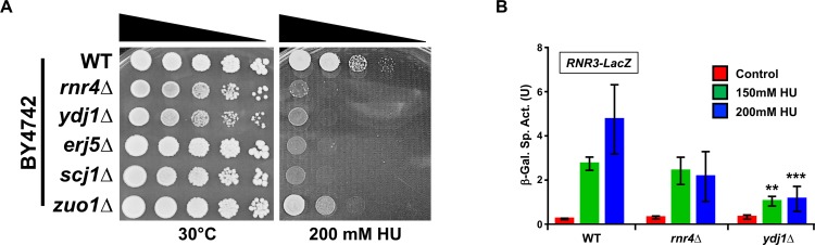
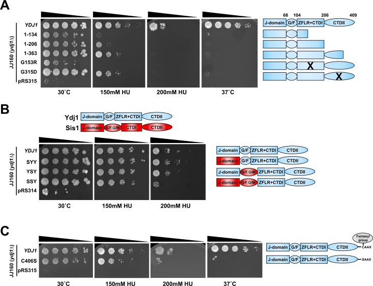
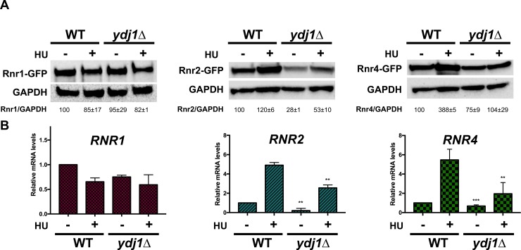
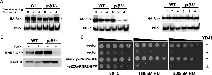
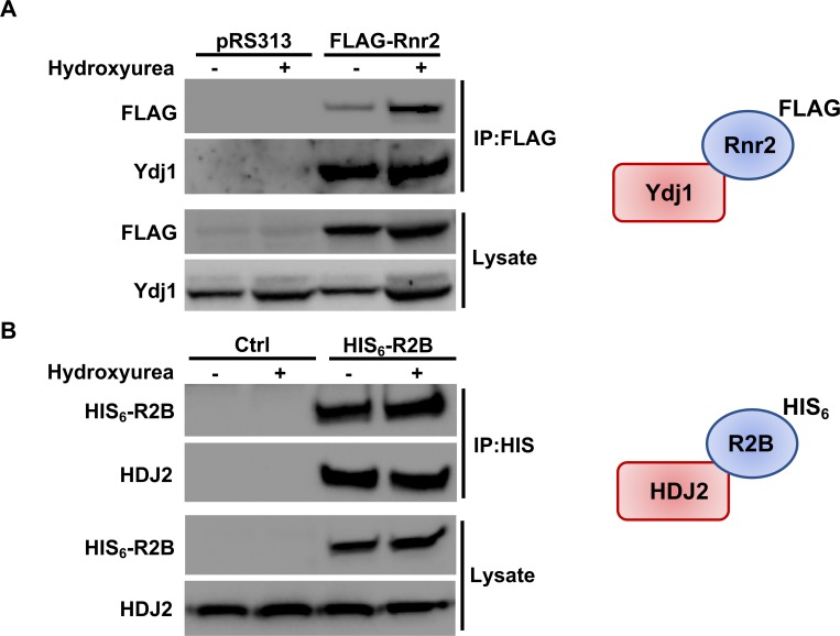
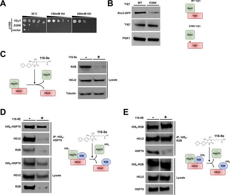
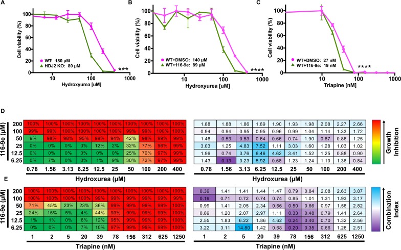
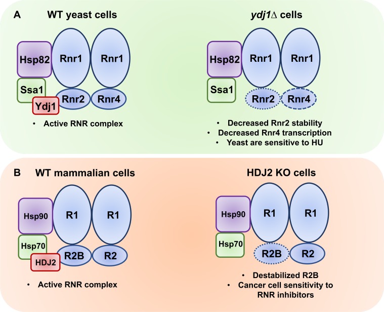
Hsp70 is a well-conserved molecular chaperone involved in the folding, stabilization, and eventual degradation of many "client" proteins. Hsp70 is regulated by a suite of co-chaperone molecules that assist in Hsp70-client interaction and stimulate the intrinsic ATPase activity of Hsp70. While previous studies have shown the anticancer target ribonucleotide reductase (RNR) is a client of Hsp70, the regulatory co-chaperones involved remain to be determined. To identify co-chaperone(s) involved in RNR activity, 28 yeast co-chaperone knockout mutants were screened for sensitivity to the RNR-perturbing agent Hydroxyurea. Ydj1, an important cytoplasmic Hsp70 co-chaperone was identified to be required for growth on HU. Ydj1 bound the RNR subunit Rnr2 and cells lacking Ydj1 showed a destabilized RNR complex. Suggesting broad conservation from yeast to human, HDJ2 binds R2B and regulates RNR stability in human cells. Perturbation of the Ssa1-Ydj1 interaction through mutation or Hsp70-HDJ2 via the small molecule 116-9e compromised RNR function, suggesting chaperone dependence of this novel role. Mammalian cells lacking HDJ2 were significantly more sensitive to RNR inhibiting drugs such as hydroxyurea, gemcitabine and triapine. Taken together, this work suggests a novel anticancer strategy-inhibition of RNR by targeting Hsp70 co-chaperone function.


Conflict of interest statement

The authors have declared that no competing interests exist.

Figures

Fig 1
Fig 1. Loss of Ydj1 impairs the DNA damage response.
(A) Yeast lacking selected Hsp70 co-chaperones are sensitive to HU. WT BY4742 or BY4742 cells lacking Rnr4, Ydj1, Erjd5, Scj1 and Zuo1 were grown overnight to saturation and serial 10-fold dilutions were plated by pin plating from 96-well plates onto YPD alone or YPD containing 200 mM HU. Plates were imaged after 3 days. (B) Cells lacking Ydj1 are compromised for DNA damage response pathway transcription. An RNR3-LacZ reporter plasmid was transformed into the indicated yeast strains. Transformants were grown and subjected to 0, 150mM or 200mM HU for 3 hours. β-Galactosidase activity was measured in crude extracts. β-Galactosidase specific activity (in units) [-Gal Sp. Act. (U)] is shown on the y axis. Each value represents the mean and standard deviation (error bar) from three independent transformants; **, P≤0.01; ***, P≤0.001 as compared to WT cell controls.
Fig 2
Fig 2. Ydj1 domains required for cellular resistance to HU.
(A) The Ydj1 C-terminus is required for HU resistance. JJ160 ydj1Δ cells transformed with plasmids encoding full-length Ydj1, truncations of Ydj1 or control plasmid pRS315 were grown to exponential phase, then 10-fold serially diluted onto media containing indicated stressor. Plates were imaged after 3 days. (B) Sis1 and Ydj1 domains are partially interchangeable for HU resistance. JJ160 ydj1Δ cells transformed with plasmids encoding full-length Ydj1 or indicated Ydj1-Sis1 fusions were grown to exponential phase, then 10-fold serially diluted onto media containing indicated stressor. Plates were imaged after 3 days. Domains of Ydj1 are colored blue, domains of Sis1 are colored red. (C) Farnesylation impacts Ydj1-mediated HU resistance. JJ160 ydj1Δ cells transformed with plasmids encoding WT Ydj1 or farnesylation-deficient mutant C406S were grown to exponential phase, then 10-fold serially diluted onto media containing indicated stressor.
Fig 3
Fig 3. RNR subunit levels in cells lacking Ydj1.
(A) BY4742 WT or ydj1Δ cells expressing endogenously tagged Rnr1-GFP, Rnr2-GFP or Rnr4-GFP were grown to exponential phase and were either left untreated or were treated with 200mM HU for 3 hours. Cell extracts were obtained, resolved on SDS-PAGE gels and analyzed by immunoblotting with anti-GFP and GAPDH antibodies. (B) Quantitation of RNR1, RNR2 and RNR4 transcription in WT or ydj1Δ cells. Levels of RNR1, RNR2 and RNR4 mRNAs in BY4742 WT or ydj1Δ cells were determined by reverse transcription and RT-qPCR. Signals of RNR1, RNR2 and RNR4 were normalized against that of ACT1 in each strain, and the resulting ratios in WT cells were arbitrarily defined as onefold. Data are the average and SD from three replicates.
Fig 4
Fig 4. Rnr2 is destabilized in ydj1Δ cells.
(A) RNR subunit stability is compromised in cells lacking Ydj1. BY4742 WT or ydj1Δ cells transformed with either pGAL1-HA-Rnr1, 2 or 4 plasmids were grown to mid-log phase in YP Galactose medium. Transcription of pGAL1-HA-Rnr1, 2 or 4 was shut off by addition of 2% glucose to cultures. Cell lysates from these samples were analyzed by Western Blotting for stability of HA-RNR subunit (HA antibody) and loading control (PGK1). (B) Examination of Rnr2 stability in WT ydj1Δ cells after translational inhibition. BY4742 WT and ydj1Δ cells expressing endogenous promoter GFP-tagged Rnr2 were grown to exponential phase in YPD media and then treated with 200 μg/ml cycloheximide for 6 hours to halt protein translation. Cell lysates were obtained and analyzed via Western Blotting for GFP-Rnr2 (GFP antibody) and a GAPDH loading control (GAPDH antibody). (C) Overexpression of Rnr2 does not suppress the HU sensitive phenotype of ydj1Δ cells. WT and ydj1Δ cells were transformed with either control plasmid pUG36 or met25p-RNR2-GFP. Transformants were grown overnight to saturation and serial 10-fold dilutions were plated by pin plating from 96-well plates onto YPD alone or YPD containing 200 mM HU. Plates were imaged after 3 days.
Fig 5
Fig 5. RNR interacts with Hsp40 in yeast and mammalian cells.
(A) Rnr2 interacts with Ydj1 in yeast. WT cells transformed with either pRS313 or plasmid expressing FLAG-tagged Rnr2 were grown to exponential phase and were either left untreated or were treated with HU as in Fig 3. Cell extracts (lysate) and immunoprecipitates (IP) with anti-FLAG M2 magnetic beads were subjected to SDS-PAGE and analyzed by immunoblotting with anti-FLAG antibodies to detect Rnr2 or anti-Ydj1 antibodies to detect Ydj1. (B) R2B interacts with HDJ2 in mammalian cells. HEK293 cells were transfected with a plasmid expressing CMV-driven HIS6-tagged R2B. Cells extracts were obtained 48 hours post-transfection. Cell extracts (lysate) and immunoprecipitates (IP) with HIS-dynabeads were subjected to SDS-PAGE and analyzed by immunoblotting with tetra-HIS antibodies to detect R2B or anti-HDJ2 antibodies to detect HDJ2.
Fig 6
Fig 6. Disruption of the Hsp70-Hsp40 interaction impacts RNR function.
(A) Mutation of the HPD motif in Ydj1 sensitizes cells to HU. ydj1Δ cells transformed with either a control plasmid, WT YDJ1 plasmid or YDJ1-D36N plasmid were grown overnight to saturation and serial 10-fold dilutions were plated by pin plating from 96-well plates onto YPD alone or YPD containing 200 mM HU. Plates were imaged after 3 days. (B) Mutation of the HPD motif in Ydj1 promotes Rnr2 degradation. ydj1Δ RNR2-GFP cells transformed with either a WT YDJ1 or YDJ1-D36N plasmid were grown to mid-log phase. Cell extracts were resolved by SDS-PAGE and analyzed by immunoblotting with anti-GFP, anti- Ydj1and anti-GAPDH antibodies. (C) Inhibition of the Hsp70-HDJ2 interaction in cancer cells promotes R2B degradation. HEK293 cells were grown to mid-confluence and then treated with 40 μM 116-9e for 72 hours. Cell extracts were subjected to SDS-PAGE and analyzed by immunoblotting with anti- R2B or anti-GAPDH antibodies. (D) 116-9E disrupts both the HSP70-HDJ2 and HSP70-R2B interaction. HEK293 cells transfected with a plasmid expressing HIS6-HSP70 were treated with 116-9E as in (C). HIS-HSP70 complexes were purified from extracts made from these cells, were subjected to SDS-PAGE and were analyzed by immunoblotting with anti-HIS, anti-R2B or anti-HDJ2 antibodies. (E) 116-9E disrupts the R2B-HSP70 interaction but leaves the R2B-HDJ2 interaction intact. HEK293 cells transfected with a plasmid expressing HIS6-R2B were treated with 116-9E as in (C). HIS-R2B complexes were purified from extracts made from these cells, subjected to SDS-PAGE and were analyzed by immunoblotting with anti-HIS, anti-R2B or anti-HDJ2 antibodies.
Fig 7
Fig 7. Inhibition of HDJ2 is synergistic with clinically-utilized RNR inhibitors.
(A) HDJ2 CRISPR KO cells are more sensitive to HU than WT cells. HAP1 WT and HDJ2 CRISPR KO cells were treated with serial dilutions of HU for 3 days. Cell viability was determined using Celltiter-Glo assay and results shown are average and SD from three replicates (***P<0.001 compared to WT cells, t-test). (B) 116-9e synergizes with HU treatment. HAP1 cells were treated with either DMSO (control) or 116-9e for 24 hours in combination with serial dilutions of HU for 3 days. Data are the average and SD from three replicates (****P<0.0001 compared to HU only treated cells, t-test). (C) 116-9e synergizes with triapine treatment. HAP1 cells were treated with either DMSO (control) or 116-9e for 24 hours, then further treated with serial dilutions of triapine for 3 days. Data are the average and SD from three replicates (***P<0.0001 compared to triapine treated cells, t-test). (D) Combination assay for 116-9e and HU. HAP1 cells were treated with combinations of 116-9e and HU for 72 h and growth inhibition was determined using CellTiter-Glo assay. Combination Index (CI, measure of drug synergy) was determined using Chou-Talalay method via Compusyn software. CI values of <1 indicate drug synergy. (E) Combination assay for 116-9e and triapine. HAP1 cells were treated with combinations of 116-9e and triapine for 72 h and growth inhibition was determined using CellTiter-Glo assay. Combination Index (CI, measure of drug synergy) was determined using Chou-Talalay method via Compusyn software. CI values of <1 indicate drug synergy.
Fig 8
Fig 8. Ydj1/HDJ2 support RNR activity in yeast and mammalian cells.
(A) In yeast, Ssa1, Hsp82 and Ydj1 bind and stabilize the RNR complex allowing dNTP synthesis required cell cycle progression and DNA repair. Loss of Ydj1 in yeast results in lowered Rnr2 levels (increased Rnr2 degradation) and Rnr4 (decreased RNR4 transcription). (B) In mammalian cells, HSP70, HSP90 and HDJ2 bind and stabilize the RNR complex allowing dNTP synthesis required cell cycle progression and DNA repair. Loss of HDJ2 activity (through either CRISPR-mediated deletion or 116-9e) promotes lowering of R2B levels, sensitizing cells to RNR-inhibiting agents such as HU and triapine.

Similar articles

Cited by

References

    1. Craig EA, Marszalek J. How Do J-Proteins Get Hsp70 to Do So Many Different Things? Trends Biochem Sci. 2017;42(5):355–68. Epub 2017/03/21. 10.1016/j.tibs.2017.02.007 ; PubMed Central PMCID: PMCPMC5409888. - DOI - PMC - PubMed
    1. Kim YE, Hipp MS, Bracher A, Hayer-Hartl M, Hartl FU. Molecular chaperone functions in protein folding and proteostasis. Annual review of biochemistry. 2013;82:323–55. Epub 2013/06/12. 10.1146/annurev-biochem-060208-092442 . - DOI - PubMed
    1. Nillegoda NB, Wentink AS, Bukau B. Protein Disaggregation in Multicellular Organisms. Trends Biochem Sci. 2018;43(4):285–300. Epub 2018/03/05. 10.1016/j.tibs.2018.02.003 . - DOI - PubMed
    1. Lianos GD, Alexiou GA, Mangano A, Mangano A, Rausei S, Boni L, et al. The role of heat shock proteins in cancer. Cancer letters. 2015;360(2):114–8. Epub 2015/02/28. 10.1016/j.canlet.2015.02.026 . - DOI - PubMed
    1. Shevtsov M, Huile G, Multhoff G. Membrane heat shock protein 70: a theranostic target for cancer therapy. Philosophical transactions of the Royal Society of London Series B, Biological sciences. 2018;373(1738). Epub 2017/12/06. 10.1098/rstb.2016.0526 ; PubMed Central PMCID: PMCPMC5717526. - DOI - PMC - PubMed

Publication types