. 2019 Mar 1;30(3):456-463.
doi: 10.1093/annonc/mdy506.

Evolutionary dynamics of residual disease in human glioblastoma

Affiliations

Evolutionary dynamics of residual disease in human glioblastoma

I Spiteri et al. Ann Oncol. .

Abstract

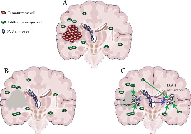
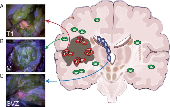
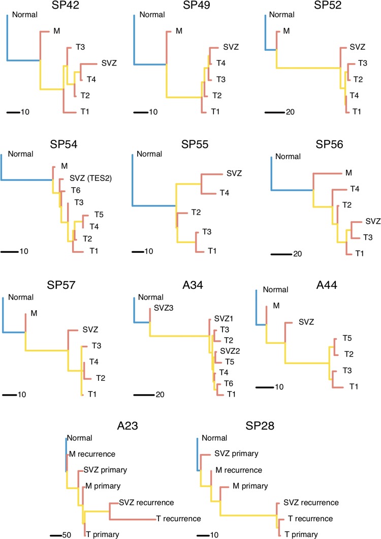
Background: Glioblastoma is the most common and aggressive adult brain malignancy against which conventional surgery and chemoradiation provide limited benefit. Even when a good treatment response is obtained, recurrence inevitably occurs either locally (∼80%) or distally (∼20%), driven by cancer clones that are often genomically distinct from those in the primary tumour. Glioblastoma cells display a characteristic infiltrative phenotype, invading the surrounding tissue and often spreading across the whole brain. Cancer cells responsible for relapse can reside in two compartments of residual disease that are left behind after treatment: the infiltrated normal brain parenchyma and the sub-ventricular zone. However, these two sources of residual disease in glioblastoma are understudied because of the difficulty in sampling these regions during surgery.

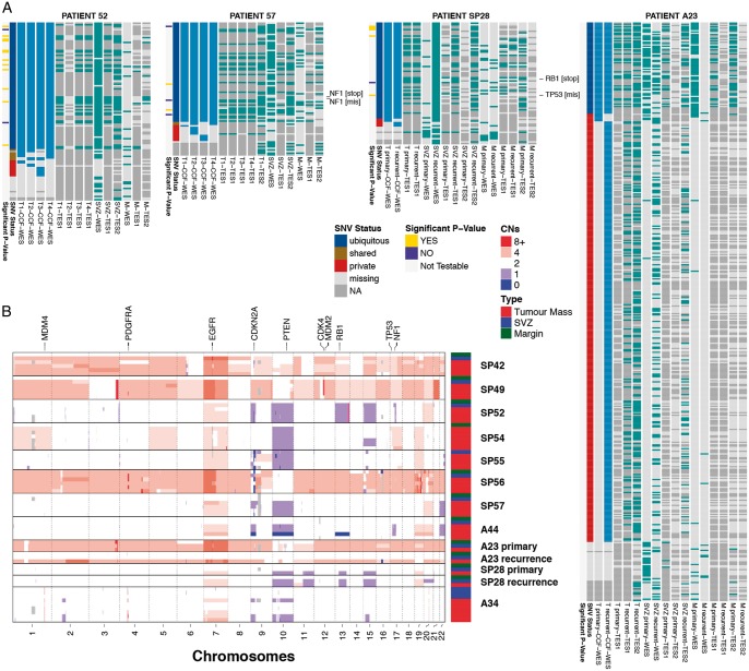
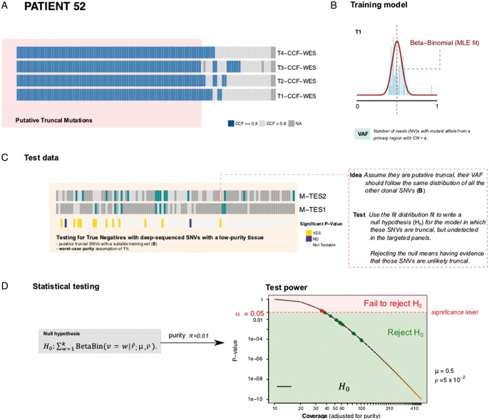
Patient and methods: Here, we present the results of whole-exome sequencing of 69 multi-region samples collected using fluorescence-guided resection from 11 patients, including the infiltrating tumour margin and the sub-ventricular zone for each patient, as well as matched blood. We used a phylogenomic approach to dissect the spatio-temporal evolution of each tumour and unveil the relation between residual disease and the main tumour mass. We also analysed two patients with paired primary-recurrence samples with matched residual disease.

Results: Our results suggest that infiltrative subclones can arise early during tumour growth in a subset of patients. After treatment, the infiltrative subclones may seed the growth of a recurrent tumour, thus representing the 'missing link' between the primary tumour and recurrent disease.

Conclusions: These results are consistent with recognised clinical phenotypic behaviour and suggest that more specific therapeutic targeting of cells in the infiltrated brain parenchyma may improve patient's outcome.

Keywords: cancer evolution; glioblastoma; phylogenetics; sub-ventricular zone; tumour margin.


Figures

Figure 1.
Figure 1.
Residual disease in glioblastoma. (A) At surgery, only the primary tumour mass (red) is removed (in dark grey the resection cavity). (B) However, infiltrative cells in the normal brain parenchyma (green) and sub-ventricular zone (SVZ) (blue) are left behind. (C) Residual glioblastoma cells infiltrated throughout the brain can give rise to relapse, both locally and distally.
Figure 2.
Figure 2.
Study design: multi-region tumour and residual disease sampling. (A) The large majority of patients present at diagnosis with a large tumour mass that is positive for 5-ALA fluorescence. In this study, we collected multiple spatially separated regions of the tumour mass (four to six regions per tumour in nine patients), as well as matched primary-relapse samples in two patients. (B) Extensive infiltration is also present in the surrounding normal brain but cancer cells are so sparse beyond the resection margin that do not appear fluorescent. Samples from the non-fluorescent infiltrative margin were collected from 9/11 patients. From paired primary-recurrent patients, we collected matched margin from the primary tumour and another margin sample from the relapsed neoplasm. (C) In a subset of patients, disease is also found in the sub-ventricular zone (SVZ), which appears fluorescent and contains malignant clones. We collected one to three samples of the SVZ from all patients, including matched SVZ in primary and relapsed tumours. Through surgery and chemo-radiation, it is possible to extensively remove the primary tumour but treatment is unlikely to completely remove the infiltrative disease, nor cancer cells in the SVZ. Those represent the majority of residual disease in glioblastoma.
Figure 3.
Figure 3.
Multi-region genomic profiles of glioblastoma residual disease. (A) For four representative patients we report the cancer cell fractions (>80%) for the tumour mass samples and presence/absence of mutation in all the residual disease samples (see supplementary Figure S1 for all cases, supplementary Table S2 for purity and supplementary data, available at Annals of Oncology online for sub-ventricular zone (SVZ) calls). Putative SNV driver events are annotated. WES, whole-exome sequencing; TES1, targeted amplicon sequencing panel 1; TES2, targeted exome capture sequencing panel 2; T1…4, tumour mass sample; SVZ, sub-ventricular zone; M, margin. (B) Digital copy number alterations are reported for each sample (see supplementary Figure S2 and Table S3, available at Annals of Oncology online for details).
Figure 4.
Figure 4.
Testing the absence of putative truncal mutations in the infiltrative margin (representative case SP52). (A) We detected single nucleotide variants (SNVs) using joint-sample variant calling from whole-exome sequencing (WES). We selected SNVs that had cancer cell fraction (CCF)0.8 in all tumour mass samples (T1, T2, …) and the same CNA status across all T samples. We call these ‘putative truncal’ SNVs. If cancer cells in M developed from T, then these mutations are ‘truly truncal’ and should be detected also in M. However, calling these mutations in the margin might be confounded by the low purity of margin samples. (B) From read counts of selected SNVs, we train for every sample a beta-binomial model of expected variant allele frequency (VAF), accounting for tumour purity and copy number status. This model describes, for each such SNV, the expected number of reads with the variant allele as a function of sample purity (i.e. we can predict how many mutant reads we expect to find in a sample like M, at purity 5%). (C) We use deep-sequencing data from targeted panels TES1 and TES2 to identify which putative truncal SNVs were not detected in the margin sample by any assay (missing SNVs). Based on the beta-binomial trained model, we created a statistical test for the null hypothesis that these mutations are truly truncal in the tumour (and hence present also in M) but remain undetected in M due to low purity. (D) Based on the expectation and the depth of coverage achieved for each tested mutation, we can calculate a P-value under the null. Rejecting the null means that we have evidence for the fact that these SNVs are not truly truncal, and that they are missing in the margin. This provides further evidence that the margin is ancestral to the tumour mass. The power of the test increases with higher coverage; we used a conservative setting of worst-case purity with πvalue (1% tumour, 99% normal) for the test, and corrected it for multiple testing via Bonferroni.
Figure 5.
Figure 5.
Phylogenetic reconstruction indicates residual disease subclones may arise early. Phylogenetic trees built with whole-exome sequencing (WES) data and excluding mutations that do not pass our test show the infiltrative margin sample at the top of the phylogeny, suggesting it contains cancer clones that occur early during tumour growth. In 6/11 samples the sub-ventricular zone (SVZ) appears as an early subclone as well. Often the phylogeny recapitulates the spatial structure of the tumour, where T1, T2, … T4 samples are taken in this order as the tumour resection extends deeper into the brain. Matched samples from M and SVZ in paired primary-relapse cases A23 and SP28 show the role of residual disease in the development of glioblastoma recurrence.

Comment in

References

    1. Stupp R, Mason WP, van den Bent MJ. et al. Radiotherapy plus concomitant and adjuvant temozolomide for glioblastoma. N Engl J Med 2005; 352(10): 987–996. - PubMed
    1. McLendon R, Friedman A, Bigner D. et al. Comprehensive genomic characterization defines human glioblastoma genes and core pathways. Nature 2008; 455(7216): 1061–1068. - PMC - PubMed
    1. Frattini V, Trifonov V, Chan JM. et al. The integrated landscape of driver genomic alterations in glioblastoma. Nat Genet 2013; 45(10): 1141–1149. - PMC - PubMed
    1. Brennan CW, Verhaak RGW, McKenna A. et al. The somatic genomic landscape of glioblastoma. Cell 2013; 155(2): 462–477. - PMC - PubMed
    1. Sottoriva A, Spiteri I, Piccirillo SGM. et al. Intratumor heterogeneity in human glioblastoma reflects cancer evolutionary dynamics. Proc Natl Acad Sci USA 2013; 110(10): 4009–4014. - PMC - PubMed

Publication types

MeSH terms

Substances