Case Reports
. 2018 Dec 1;27(23):4135-4144.
doi: 10.1093/hmg/ddy305.

Mutations of the mitochondrial carrier translocase channel subunit TIM22 cause early-onset mitochondrial myopathy

Affiliations
Case Reports

Mutations of the mitochondrial carrier translocase channel subunit TIM22 cause early-onset mitochondrial myopathy

David Pacheu-Grau et al. Hum Mol Genet. .

Abstract

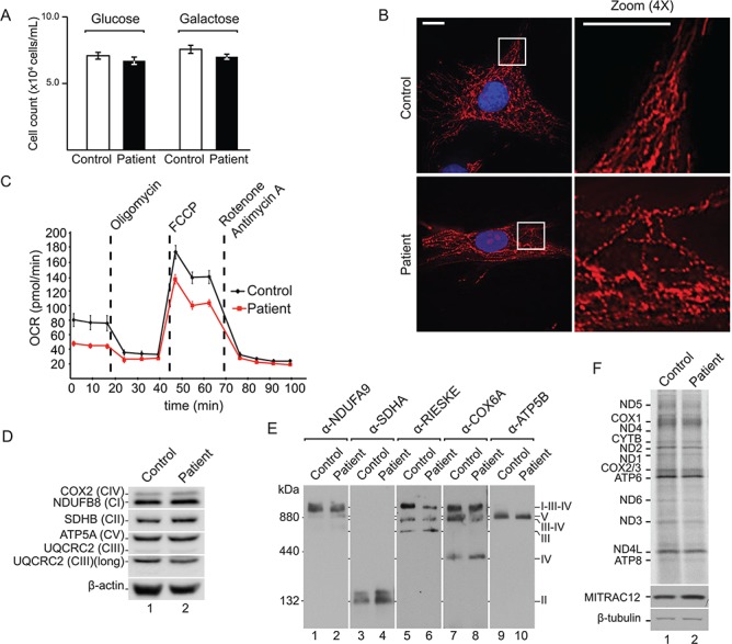
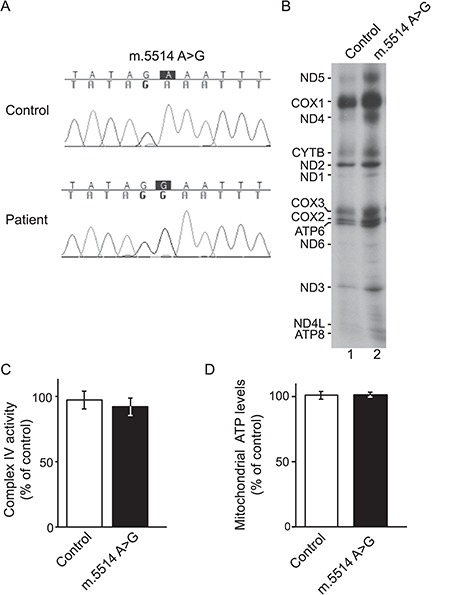
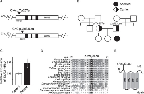
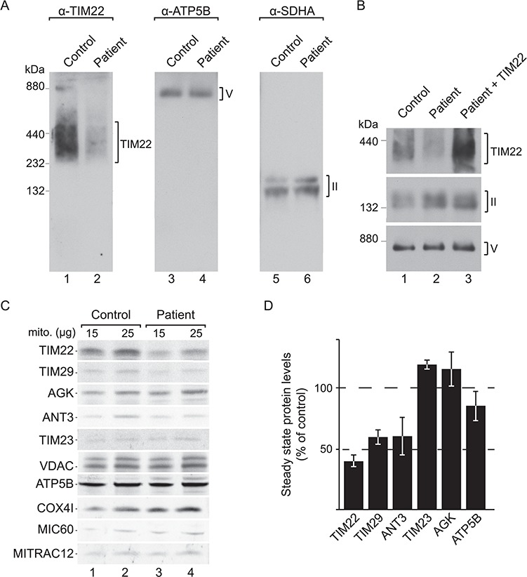
Protein import into mitochondria is facilitated by translocases within the outer and the inner mitochondrial membranes that are dedicated to a highly specific subset of client proteins. The mitochondrial carrier translocase (TIM22 complex) inserts multispanning proteins, such as mitochondrial metabolite carriers and translocase subunits (TIM23, TIM17A/B and TIM22), into the inner mitochondrial membrane. Both types of substrates are essential for mitochondrial metabolic function and biogenesis. Here, we report on a subject, diagnosed at 1.5 years, with a neuromuscular presentation, comprising hypotonia, gastroesophageal reflux disease and persistently elevated serum and Cerebrospinal fluid lactate (CSF). Patient fibroblasts displayed reduced oxidative capacity and altered mitochondrial morphology. Using trans-mitochondrial cybrid cell lines, we excluded a candidate variant in mitochondrial DNA as causative of these effects. Whole-exome sequencing identified compound heterozygous variants in the TIM22 gene (NM_013337), resulting in premature truncation in one allele (p.Tyr25Ter) and a point mutation in a conserved residue (p.Val33Leu), within the intermembrane space region, of the TIM22 protein in the second allele. Although mRNA transcripts of TIM22 were elevated, biochemical analyses revealed lower levels of TIM22 protein and an even greater deficiency of TIM22 complex formation. In agreement with a defect in carrier translocase function, carrier protein amounts in the inner membrane were found to be reduced. This is the first report of pathogenic variants in the TIM22 pore-forming subunit of the carrier translocase affecting the biogenesis of inner mitochondrial membrane proteins critical for metabolite exchange.


Figures

Figure 1
Figure 1
Composition of human TIM22 complex and mechanism of action. The TIM22 complex is comprised of the central twin-pore forming unit TIM22, in addition to TIM10B and the metazoan specific subunits, TIM29 and AGK. A soluble hexameric ring, made up of TIM9 and TIM10A and present in the IMS, guides precursors (carrier proteins or TIM22, TIM23, TIM17A/B) from the TOM complex to the TIM22 complex. Upon docking to the TIM22 complex, the precursor is released into the TIM22 channel and its insertion into the membrane is driven by the mitochondrial membrane potential Δψ.
Figure 2
Figure 2
Patient fibroblasts exhibit reduced mitochondrial fitness. (A) Cell counts of control and patient cells after 3 days of growth on glucose or galactose (Standard error of the mean (SEM), n = 3). (B) Mitochondrial network of a representative control and patient fibroblast cell, stained with MitoTracker and DAPI. Scale bar = 15 μm. (C) Real-time respirometry of patient and control cells; OCR. (D) SDS-PAGE and western blot analysis of OXPHOS subunits with indicated antibodies in control and patient fibroblast lysates using β-actin as loading control. (E) blue native polyacrylamide gel electrophoresis (BN-PAGE) and western blot analysis of OXPHOS complexes in solubilized mitochondria from control and patient fibroblasts using indicated antibodies. (F) In vivo labelling of mitochondrial translation products in control and patient immortalized cells. Samples were analysed by SDS-PAGE, digital autoradiography and western blotting.
Figure 3
Figure 3
A nuclear genetic basis of the mitochondrial phenotype. (A) Sequencing electropherogram showing the presence or absence of the m.5514A>G in A549 cybrid cells. (B) In vivo labelling of mitochondrial translation products in control and mutant cybrids. Samples were analysed by SDS-PAGE and autoradiography. (C) Cytochrome c oxidase enzymatic activity, normalized by the enzymatic activity of CS, of mutant cybrid cells relative to control cybrids (Standard deviation (STDEV), n = 3). (D) Levels of mitochondrial ATP in mutant cybrids compared to control (STDEV, n = 6).
Figure 4
Figure 4
Identification of compound heterozygous variants in TIM22. (A) New mutations identified in the TIM22 gene locus. (B) Family pedigree showing patient and known unaffected relatives carrying single heterozygous variants. Circle denotes female and square denotes male family members. (C) Quantitative PCR of TIM22 mRNA levels in patient fibroblasts, relative to control fibroblasts (set to 1.0). TIM22 mRNA levels were normalized to HPRT (SEM, n = 3). (D) Amino acid sequence alignment of the p.Val33Leu containing-segment of TIM22 in indicated organisms. (E) Position of variant p.Val33Leu according to the predicted topology of TIM22.
Figure 5
Figure 5
The p.Val33Leu variant destabilizes the carrier translocase and affects metabolite carrier levels. (A) Isolated mitochondria from control and patient fibroblasts were solubilized and resolved using BN-PAGE, followed by western blotting and detection using the indicated antibodies. (B) Patient immortalized fibroblast cells were electroporated with wild-type TIM22. Mitochondria from these cells and from non-electroporated control and patient cells were isolated, solubilized and analysed by BN-PAGE, followed by western blotting and detection using the indicated antibodies. (C) Isolated mitochondria from control and patient fibroblasts were subjected to SDS-PAGE and western blotting. (D) Quantification of western blotting signals using infrared secondary antibodies in patient mitochondria compared to control and normalized to the VDAC signal (SEM, n = 3).

References

    1. Dudek J., Rehling P. and Laan M. (2013) Mitochondrial protein import; common principles and physiological networks. Biochim. Biophys. Acta, 1833, 274–285. - PubMed
    1. Wiedemann N. and Pfanner N. (2017) Mitochondrial machineries for protein import and assembly. Annu. Rev. Biochem., 86, 685–714. - PubMed
    1. Endo T., Yamano K. and Kawano S. (2011) Structural insight into the mitochondrial protein import system. Biochim. Biophys. Acta, 1808, 955–970. - PubMed
    1. Neupert W. and Herrmann J.M. (2007) Translocation of proteins into mitochondria. Annu. Rev. Biochem., 76, 723–749. - PubMed
    1. Chacinska A., Koehler C.M., Milenkovic D., Lithgow T. and Pfanner N. (2009) Importing mitochondrial proteins; machineries and mechanisms. Cell, 138, 628–644. - PMC - PubMed

Publication types

MeSH terms

Substances