Review
. 2019 May;139(5):1118-1126.
doi: 10.1016/j.jid.2018.10.038. Epub 2018 Nov 16.

Collagen Prolyl Hydroxylases Are Bifunctional Growth Regulators in Melanoma

Affiliations
Review

Collagen Prolyl Hydroxylases Are Bifunctional Growth Regulators in Melanoma

Aithne Atkinson et al. J Invest Dermatol. 2019 May.

Abstract

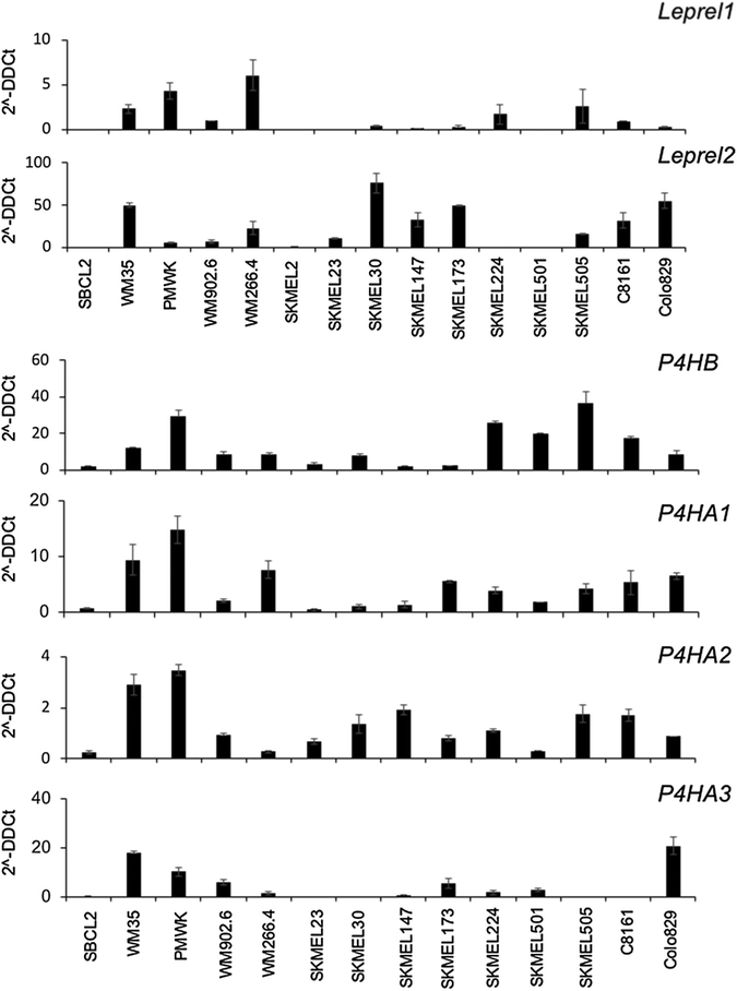
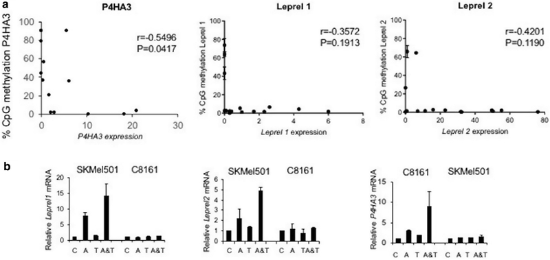
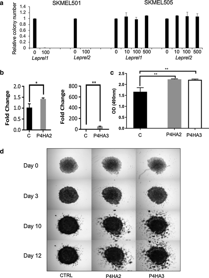
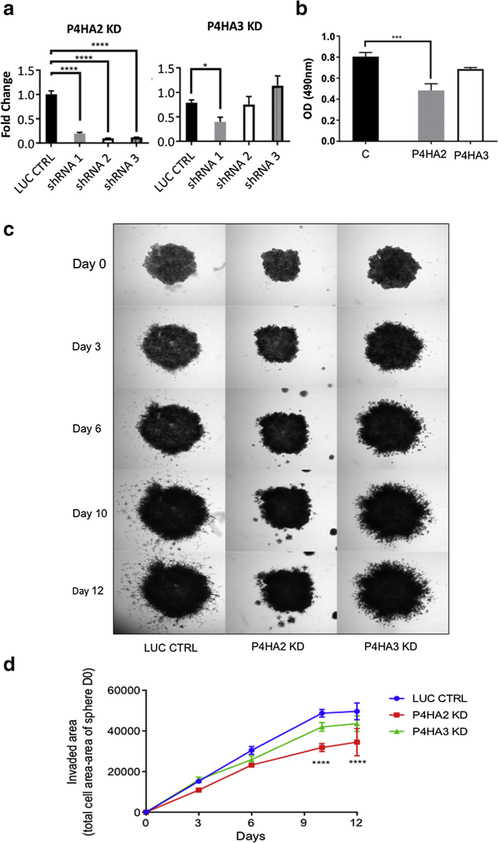
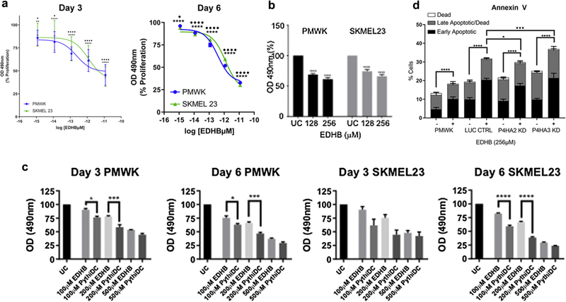
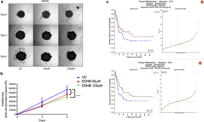
Appropriate post-translational processing of collagen requires prolyl hydroxylation, catalyzed by collagen prolyl 3-hydroxylase and collagen prolyl 4-hydroxylase, and is essential for normal cell function. Here we have investigated the expression, transcriptional regulation, and function of the collagen prolyl 3-hydroxylase and collagen prolyl 4-hydroxylase families in melanoma. We show that the collagen prolyl 3-hydroxylase family exemplified by Leprel1 and Leprel2 is subject to methylation-dependent transcriptional silencing in primary and metastatic melanoma consistent with a tumor suppressor function. In contrast, although there is transcriptional silencing of P4HA3 in a subset of melanomas, the collagen prolyl 4-hydroxylase family members P4HA1, P4HA2, and P4HA3 are often overexpressed in melanoma, expression being prognostic of worse clinical outcomes. Consistent with tumor suppressor function, ectopic expression of Leprel1 and Leprel2 inhibits melanoma proliferation, whereas P4HA2 and P4HA3 increase proliferation, and particularly invasiveness, of melanoma cells. Pharmacological inhibition with multiple selective collagen prolyl 4-hydroxylase inhibitors reduces proliferation and inhibits invasiveness of melanoma cells. Together, our data identify the collagen prolyl 3-hydroxylase and collagen prolyl 4-hydroxylase families as potentially important regulators of melanoma growth and invasiveness and suggest that selective inhibition of collagen prolyl 4-hydroxylase is an attractive strategy to reduce the invasive properties of melanoma cells.


Conflict of interest statement

CONFLICT OF INTEREST

The authors state no conflict of interest.

Figures

Figure 1.
Figure 1.. Expression of C-P3H and C-P4H genes in melanoma cell lines.
Quantitative PCR analysis of each C-P3H and C-P4H genes in the melanoma cell line panel. cDNA was prepared and quantitative PCR performed as described in Materials and Methods. Expression is shown as 2-ΔΔCt calculated as described in Materials and Methods. Data shown are mean ± 1 standard deviation. Each experiment was performed in triplicate and repeated at least twice.
Figure 2.
Figure 2.. C-P3H and C-P4H are epigenetically regulated in melanoma.
(a) Scatter plot showing that methylation in the P4HA3, Leprel1 and Leprel2 CpG islands is negatively correlated with expression. Data are mean ± standard error of the mean (n = 15). Correlation analysis was done using Pearson’s correlation coefficient. (b) Demethylation reactivates expression of P4HA3 in melanoma cells with CpG island methylation. C8161 and SKMEL501 cells were grown in the presence of azacytidine (A), trichostatin A (T) or both agents (A&T). Expression was determined by quantitative PCR. Data shown are mean ± 1 standard deviation relative to control cells (C).
Figure 3.
Figure 3.. Collagen prolyl 3-hydroxylase and collagen prolyl 4-hydroxylase have opposing effects on melanoma progression.
(a) Ectopic expression of Leprel1 and Leprel2 in cells lacking endogenous expression blocks proliferation. (b) Quantitative PCR analysis in PMWK engineered to express of P4HA2 and P4HA3. Data are mean ± standard error of the mean (n = 3). Significance was tested using one-way analysis of variance with Dunnett’s post-hoc testing.(c) P4HA2 and P4HA3 overexpression significantly increase proliferation in PMWK cells. Graphs shows OD at 490 nm 6 days post treatment. Data are mean ± standard error of the mean over biological repeats (n = 2) performed in triplicate. Significance was tested using two-way analysis of variance with Tukey’s post-hoc testing. (d) P4HA2 and P4HA3 overexpression increase the invasiveness relative to control (CTRL) of PMWK early radial growth phase melanoma cells. Images are representative of two independent experiments performed in triplicate. OD, optical density.
Figure 4.
Figure 4.. Inhibition of collagen prolyl 4-hydroxylase gene expression reduces melanoma proliferation and invasiveness.
(a) Quantitative PCR analysis of P4HA2 and P4HA3 in PMWK cells expressing short hairpin RNA. Data are mean 2-ΔΔCt ± standard error of the mean (n = 3). Significance: one-way analysis of variance with Dunnett’s post-hoc testing. (b) P4HA2 and P4HA3 knockdown reduce proliferation of PMWK cells. Quantification was performed using sulforhodamine b as described in Materials and Methods. Data are mean ± standard error of the mean over biological repeats (n = 2) performed in triplicate. Significance shows the difference to appropriate control, using one-way analysis of variance with Sidak’s post-hoc testing.(c) Collagen prolyl 4-hydroxylase knockdown reduces invasiveness of PMWK cells. Representative images are shown. Spheroids were imaged at ×2.(d) Reduced invasion of PMWK 3-dimensionl spheroids. Data are mean ± standard error of the mean (n = 2), in triplicate. Significance: two-way analysis of variance with Tukey’s post-hoc testing. KD, knockdown; LUC CTRL, luciferase control; shRNA, short hairpin RNA.
Figure 5.
Figure 5.. Inhibition of collagen prolyl 4-hydroxylase has antiproliferative effects on melanoma cells via induction of apoptosis.
(a, b) Inhibition of proliferation of PMWK (blue) and SKMEL23 (green) by EDHB. Data are mean ± standard error of the mean over biological repeats (n = 3) performed in six replicates. Significance: two-way analysis of variance with Tukey’s post-hoc testing. (c) Antiproliferative effect of EDBH and PythiDC on PMWK and SKMEL23. Data are mean ± standard error of the mean (n = 2) in triplicate. Significance: one-way analysis of variance with Sidak’s post-hoc testing. (d) EDHB induces apoptosis in PMWK cells. Apoptosis was measured via Annexin V staining on day 3. Data are mean ± standard error of the mean (n = 3). Live cell population not shown. Significance: two-way analysis of variance with Tukey’s post-hoc testing. *Differences in early-stage apoptosis. Differences in late-stage apoptosis. EDHB, ethyl dihydroxybennzoic acid; PythiDC, 2-(5-carboxythiazol-2-yl)pyridine-5-carboxylic acid.
Figure 6.
Figure 6.. Blocking collagen prolyl 4-hydroxylase inhibits melanoma invasion.
(a) EDHB inhibits invasiveness of PMWK 3-dimensional tumor spheroids. Representative images from two independent experiments performed in triplicate. Spheroids were imaged at ×2 on days 0, 3, and 6 post treatment.(b) Quantification of the effect of EDHB on PMWK 3-dimensional spheroid invasion. Spheroids were imaged at ×2 on days 0, 3, and 6 post treatment. Data are mean ± standard error of the mean (n = 2) performed in triplicate. Significance was compared to the appropriate control using two-way analysis of variance with Tukey’s post-hoc testing. (c) Overexpression of C-P4H genes is associated with worse disease-free survival in melanoma. Kaplane–Meier plots by gene expression were created using the “Tumor Melanoma-Jönsson-214-custom-ilmnht12v4” data set (n = 214) (Cirenajwis et al., 2015). EDHB, ethyl dihydroxybennzoic acid.

References

    1. Cirenajwis H, Ekedahl H, Lauss M, Harbst K, Carneiro A, Enoksson J, et al. Molecular stratification of metastatic melanoma using gene expression profiling: prediction of survival outcome and benefit from molecular targeted therapy. Oncotarget 2015;6:12297–309. - PMC - PubMed
    1. Fernandes RJ, Farnand AW, Traeger GR, Weis MA, Eyre DR. A role for prolyl 3-hydroxylase in post translational modification of fibril-forming collagens. J Biol Chem 2011;286:30662–9. - PMC - PubMed
    1. Gilkes DM, Chatuverdi P, Bajpai S, Wong CC, Wei HH, Pitcairn S, et al. Collagen prolyl hydroxylases are essential for breast cancer metastasis. Cancer Res 2013;73:3285–96. - PMC - PubMed
    1. Gorres KL, Raines RT. Prolyl 4-hydroxylase. Crit Rev Biochem Mol Biol 2010;45:106–24. - PMC - PubMed
    1. Hatzimichael E, Lo Nigro C, Lattanzio L, Syed N, Shah R, Dasoula A, et al. The collagen prolyl hydroxylases are novel transcriptionally silenced genes in lymphoma. Br J Cancer 2012;107:1423–32. - PMC - PubMed

Publication types

MeSH terms