

doi: 10.1016/j.jaci.2018.10.053.
Epub 2018 Nov 17.
Role of P2X3 receptors in scratching behavior in mouse models
Affiliations
- PMID: 30452925
- PMCID: PMC7092727
- DOI: 10.1016/j.jaci.2018.10.053
Item in Clipboard
Role of P2X3 receptors in scratching behavior in mouse models
J Allergy Clin Immunol.
2019 Mar.
No abstract available
Conflict of interest statement
Disclosure of potential conflict of interest: The authors declare that they have no relevant conflicts of interest.
Figures
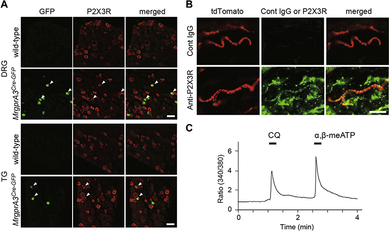
MrgprA3-positive sensory neurons express functional P2X3Rs. A, Immunofluorescence images of DRG/TG neurons of wild-type and MrgprA3GFP-Cre mice. The MrgprA3+ neurons (GFP+ cells, green) were counterstained for P2X3R immunoreactivity (red). Arrowheads indicate double-positive neurons. B, Immunofluorescence images of the skin of Mrgpra3GFP-Cre;ROSA26tdTomato mice. MrgprA3+ fibers (tdTomato+, red) were counterstained for P2X3R (bottom, green) or isotype control (Cont IgG, top, green). Scale bar, 20 μm. C, Representative intracellular Ca2+ responses to chloroquine (CQ) and α,β-meATP in DRG neurons.
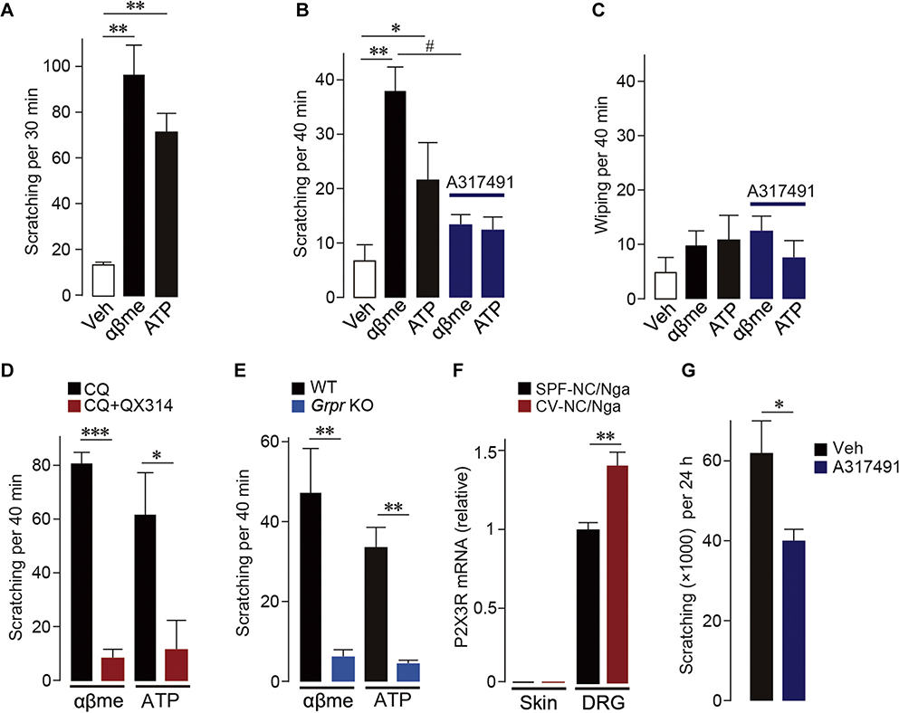
Activation of P2X3Rs in MrgprA3-positive sensory neurons induces scratching behavior mediated via GRPR signaling. A, Scratching induced after intradermal injection of ATP and α,β-meATP (αβme) into the rostral back (n = 6). B and C, Scratching (Fig 2, B) or wiping (Fig 2, C) induced after cheek injection of ATP and α,β-meATP (αβme) with or without A317491, a selective antagonist for P2X3Rs (n = 6–10). D, Effect of coinjection of chloroquine (CQ) and QX-314 on ATP and α,β-meATP (αβme)-induced scratching (n = 5–6). E, Scratching evoked by ATP and α,β-meATP (αβme) in Grpr-knockout (KO) mice (n = 6–7). F, P2X3R mRNA in the skin or DRG of SPF (control, maintained in specific pathogen-free condition) or CV-NC/Nga mice (n = 4). G, Effect of intradermal injection of A317491 on scratching in CV-NC/Nga mice (n = 7–9). Veh, Vehicle; WT, Wild-type. Data are shown as means ± SEM. Mann-Whitney U test (Fig 2, A, E, and G), unpaired t test (Fig 2, D and F), and 1-way ANOVA followed by post hoc Tukey multiple comparisons test (Fig 2, B and C) were used for statistical analyses. *,#P < .05, **P < .01, ***P < .001.

P2X3R mRNA expression in the skin, DRG, and TG. P2X3R mRNA in the skin, cervical DRG, or TG of C57BL/6J mice (n = 3). Values represent the relative ratio of P2X3R mRNA (normalized to GAPDH mRNA value) to the value of the DRG. P2X3R mRNA expression levels in the skin were very low compared with those in the DRG (<0.02%). GAPDH, Glyceraldehyde 3-phosphate dehydrogenase. Data are shown as means ± SEM.

Expression profiles of P2X3R and MrgprA3+ neurons. The Venn diagram illustrating the expression profiles of DRG neurons positive for P2X3R, MrgprA3, and/or TRPV1 in MrgprA3Cre-GFP mice. The sizes of the circles represent the proportion of each neuronal population.

Dose-dependent increase in scratching induced by P2X3R agonists. Dose-responses for increase in scratching after cheek injection of ATP or α,β-meATP (n = 4–6). Veh, Vehicle. Data are shown as means ± SEM. *P < .05, unpaired t test; **P < .01, Mann-Whitney U test.

Effect of A317491 on scratching caused by intradermal injection of α,β-meATP into the rostral back. α,β-meATP (αβme: 200 μg/50 μL) was intradermally injected into the rostral back with or without A317491 (A31: 200 μg/50 μL), a selective antagonist for P2X3Rs (n = 6). Scratching behavior was counted for 30 minutes after α,β-meATP injection. Data are shown as means ± SEM. *P < .05, unpaired t test.</_Caption>

Effect of A317491 on skin inflammation of NC/Nga mice. A, Photographs of hematoxylin-eosin–stained deparaffinized sections of the rostral back skin taken from SPF (control) and CV (chronic scratching)-NC/Nga mice 24 hours after intradermal administration of vehicle (Veh) or A317491 (A31). Scale bar, 50 μm. B, Number of inflammatory cells in the skin of SPF, CV (Veh), and CV (A31)-NC/Nga mice (n = 3). C, Epidermal thickness of SPF, CV (Veh), and CV (A31)-NC/Nga mice (n = 3). SPF, Specific pathogen free. Data are shown as means ± SEM.
References
-
- Miller G Grasping for clues to the biology of itch. Science 2007;318:188–9. - PubMed
Publication types
MeSH terms
Substances
Grants and funding
LinkOut - more resources
Full Text Sources
Other Literature Sources
Medical
